Fibrosis
As fibrosis (fachsprachlich also Fibrosis ) is a pathological increase of connective tissue, referred to in human and animal tissues and organs, whose main component is collagen fibers are. The tissue of the affected organ is hardened. Scarred changes develop which, in an advanced stage, lead to a restriction of the respective organ function. Substances that cause fibrosis are called fibrogens , the associated adjective is fibrogenic .
etiology
There are so-called primary fibroses in which the cause of the change is unknown. The respective tissue fibrosis without any noticeable external damage. An example of primary fibrosis is retroperitoneal fibrosis Ormond's disease . Secondary fibroses, however, are far more common . In these, the tissue is initially caused by an exogenous , i.e. externally acting, or endogenous , i.e. by the body itself, damage, for example through inflammation or circulatory disorders. Such damage is called a noxious agent . This activates special cells within the connective tissue, so-called fibroblasts , which increasingly produce interstitial connective tissue within the destroyed tissue . The connective tissue that runs between individual organ sections within the organs is called interstitial. The blood vessels and nerves that supply the organs are located in it. Therefore, the fibrosis, i.e. the uncontrolled growth of these areas that permeate the organs, causes scarring that can severely impair the functionality of the affected organs or, in extreme cases, destroy it.
In principle, this process is possible in any tissue. The best-known example is alcohol-toxic liver cirrhosis , in which fibrosis destroys the liver parenchyma , i.e. the pollutant-degrading areas of the liver, through repeated and long-lasting poisoning with alcohol. Further examples of diseases caused by fibrosis are pulmonary fibrosis , the loss of function of the kidneys after long-term chronic renal insufficiency or diastolic dysfunction of the heart , as the end stage of the most varied of lung diseases .
Affected organs
Postoperative connective tissue overgrowth can trigger fibrosis in the eye. As a result of endocrine orbitopathy, there is a noticeable protrusion of the eyes, raised upper eyelids and widening of the eyelid fissures .
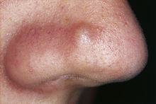
Fibrosis can cause a keloid on the skin . Dupuytren's disease can occur on the palms of the hands . The induratio penis plastica is responsible for a connective tissue disease of the penis , which can lead to penile curvature , pain during an erection and even erectile dysfunction with mental disorders. The fibrosis can form a hamartoma on the nasal papule, which is called a fibrous nasal papule .
The cardiac arrhythmia atrial fibrillation is possible. It is associated with an increased risk of stroke and heart failure . Heart muscle disease ( cardiomyopathy ) can be another symptom of cardiac fibrosis. One is also diastolic heart failure , a myocardial infarction or remodeling with fibrosis at the heart possible. An increase in connective tissue also takes place in diabetic cardiomyopathy .
The fibrosis can also affect the lungs, which are then classified as pulmonary fibrosis . The lungs, along with the skin, can be affected as a result of scleroderma . A special syndrome of scleroderma is the CREST syndrome . Asthma is also thought to be the result of pulmonary fibrosis.
In the kidney, fibrosis can cause chronic kidney failure . A peritoneal dialysis is used to treat a blood purification procedure. In diabetic nephropathy , the connective tissue also increases. People with a kidney transplant are at risk of chronic transplant nephropathy , which is the most common cause of premature loss of function of a transplanted kidney.
A fatty liver is a potential target steatohepatitis in liver fibrosis. In the final stage this leads to liver cirrhosis , which among other things causes circulatory disorders in the liver.
Fibrosis in the stomach area can cause inflammation of the lining of the large intestine ( microscopic colitis ). The retroperitoneal fibrosis allows the connective tissue between the rear peritoneum and the spine with encasement of the vessels, nerves and the ureter multiply.
In osteomyelofibrosis , the blood-forming bone marrow is affected. It can lead to anemia or thrombocytopenia . The secondary version of fibrosis can be caused by radiation therapy.
The drug ciclosporin , which transplant patients must take, can lead to a fibroadenoma in high doses .
Pathogenesis
The cells are divided into epithelial cells and mesenchymal cells . An epithelium consists of a basement membrane that forms the base and the epithelial cells on top.
What all epithelial cells have in common is their polarity : They have an outer side called apically opposite the basal underside, which faces the basement membrane. These have different functions. Epithelia are arranged close to one another and thus form a cover or glandular tissue in the form of an interface that carries out specialized metabolic functions. In the kidney, for example, the epithelium is located in the tubule , a fine tube in which the urine flowing through is concentrated. The transporting epithelium absorbs certain substances and secretes others. It is renewed by dividing stem cells that are occasionally located on the basement membrane.
A mesenchymal cell, on the other hand, is typically not bound to a basement membrane, but is usually freely mobile like the cells of the immune system. It can move in the bloodstream and pass through tissues.
The fibroblasts are a form of mesenchymal cells. They fill the space between the cells with an extracellular matrix of a small structure and, as a kind of scar tissue, complement damaged areas of the epithelium.
When fibrosis develops, differentiated epithelial cells take on the properties of a mesenchymal cell. They turn into myofibroblasts . These are a special form of fibroblasts that are normally responsible for wound healing, among other things. This transformation process is called epithelial-mesenchymal transition .
The myofibroblasts are very productive, they generate far more matrix material than would be required and thus considerably impair the function of the cells surrounding them.
The bone marrow forms mesenchymal progenitor cells, so-called fibrocytes , which can migrate into the organs and become fibroblasts.
Three factors influence this process: TGFbeta , p38- MAPK and thrombospondin . The thrombospondin is antiangiogenic ; it reduces the ingrowth of blood vessels into the scarring areas and thus causes a lack of energy in the adjacent epithelia. In the case of liver cirrhosis, the reduced conductivity results in an increased perfusion resistance , so that bypass circuits are formed.
Organ-specific fibroses of the liver, lungs, kidneys or heart, which lead to functional impairment, are clinically evident. In the area of the rheumatic entities , fibrosis can develop that affect several organs, such as scleroderma , which in addition to thickening of the skin also changes the lungs and small kidney vessel walls.
therapy
The most radical form of therapy is to destroy the myofibroblasts. So-called NK cells are used for this. NK cells normally destroy the body's own cells that are infected by viruses or other attackers. However, the most practical form of therapy is to inhibit signal transduction of the signaling molecule TGFbeta somewhere . Many lead and old substances are also known which, for example, inhibit p38- MAPK . Substances that have been recognized to lead to TGFbeta activation, such as angiotensin or aldosterone , are influenced by drugs available today.
literature
- D. Reinhardt, M. Götz, R. Kraemer, M. Schöni: Cystic Fibrosis. Springer-Verlag, 2013 ( online )
Web links
- Fibrosis in the DocCheck Lexicon
- Fibrosis: causes, symptoms and therapy on hiv-symptome.de
- Fibrosis on onmeda.de
- Fibrosis on www.healthpedia.de
- Fibrosis on symptomat.de
- Arnd Petry: Fibrosis paralyzes the organs with scars. In: Die Welt, 12 February 2015
