Dupuytren's disease
| Classification according to ICD-10 | |
|---|---|
| M72.0 | Fibromatosis of the palmar fascia [Dupuytren's contracture] |
| ICD-10 online (WHO version 2019) | |
The Dupuytren's contracture ( Dupuytren's contracture , IPA pronunciation: [dypɥ'tʁɑ]) is a benign disease of the connective tissue of the palm ( palmar ). In 1832 Baron Guillaume Dupuytren (1777-1835) presented the disease named after him in Paris. However, it was first described by Henry Cline and Sir Astley Cooper in 1777. The triggering cause could not be found until today. Dupuytren's disease belongs to the group of fibromatoses . The ring or little finger is usually affected, but any finger can be affected.
Epidemiology
Dupuytren's disease usually occurs in middle age, 85% of the affected patients are men, in whom the disease typically occurs earlier than in women. The Dupuytren's contracture occurs predominantly in Central and Northern Europe and North America, but less often in Africa and Asia. More recently, some cases have also been reported from Japan and China. The prevalence is very variable and ranges from 2 to 42% in the western industrialized countries. The condition is more common with age. It is associated with alcohol-toxic liver damage and tobacco smoking as well as diabetes mellitus . First and foremost there is a strong familial accumulation. Typically, small and ring finger affected contractures are found almost exclusively on the Fingergrund- and -mittelgelenken . In half of the cases, the Dupuytren muscle is found on both hands; overall, the right hand is affected slightly more often. A genetic component is considered certain; every third person affected has a family member also affected by the disease.
Symptoms and course
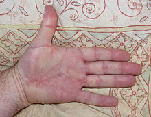
Characteristic of the disease is the appearance of knots and strands on the inner surface of the hand.
The natural course is variable and progression is often slow over years. However, a standstill is also possible at any stage. It often takes years before the initially palpable, nodular or cord-like hardening leads to a restriction of mobility in the metacarpophalangeal and median joints of the fingers (flexion contracture). As a rule, treatment, namely an operation, is only considered at this advanced stage.
The thumb is the third most affected after the ring and little fingers. Studies show a participation between 3 and 28%. This leads to painful knots between the first and second metacarpal bones and in the area of the thenar (ball of the thumb). Above all, extension and abduction of the thumb are restricted.
Fibromatoses
Similar and well-related diseases are Ledderhose's disease , i. H. the corresponding disease on the inside of the feet (plantar aponeurosis), which is found in 5–20% of patients with Dupuytren 's disease , and possibly penile curvature (in 4%), also known as Peyronie's disease or induratio penis plastica . Occasionally, symptomatically similar growths on the abdominal wall (fasciitis nodularis) are associated with Dupuytren's disease, as well as retroperitoneal fibrosis Ormond's disease.
These diseases are often summed up as fibromatoses , which are benign tumor-like connective tissue growths that can often grow aggressively. The growths originate from myofibroblasts, and in addition to genetic factors, disorders of the estrogen metabolism are also discussed as causes. In 2011, various changes in the Wnt signaling pathway were linked to the occurrence of the disease.
The WHO classifies palmar and plantar fibromatosis as an independent entity “superficial fibromatoses” (superficial fibromatoses) in the large group of fibroblastic and myofibroblastic tumors.
Degrees of severity
The Tubiana classification - according to Raoul Tubiana (1915–2013) - in six degrees of severity is common. In stage 0 there is no lesion. The degree N (for nodus = knot or nodulus = nodule) describes the cord or knots in the palm of the hand without a stretch deficit. For the division into one of the four following stages (separately for both hands), the sum of the extension deficits of all joints of the most affected finger is calculated. You can also specify this sum individually for each affected finger.
- Grade 1: Sum of the extension deficits 0 ° to 45 °
- Grade 2: Sum of the extension deficits 45 ° to 90 °
- Grade 3: Sum of the extension deficits 90 ° to 135 °
- Grade 4: Sum of the extension deficits 135 ° to 270 °
According to the laws of geometry , this sum of angles is identical to the deviation of the end link from the horizontal starting position. For example, if the extension deficits of the affected little finger are 45 ° in the base joint, 100 ° in the middle joint and 15 ° in the end joint, then the sum of the angles is 160 °. This angle sum of 160 ° with stage 4 results solely from the misalignment of the end link. A maximum of about 270 ° is achieved with right-angled stiffeners in all three finger joints; here the three phalanxes form a square together with the metacarpal bone . Sometimes stage 1 is divided into two stages: stage N / 1 with an angle sum between 0 ° and 5 ° and degree 1 with a sum between 6 ° and 45 °.
Occasionally there is also a division of Dupuytren's disease into only three levels (light, medium and severe involvement of one hand). There is also an older classification according to Henry William Meyerding (1884–1969):
- Stage I: Knot formation without further restrictions on extension and spreadability
- Stage II: indurations with slight flexion in the base joint
- Stage III: involvement of the middle phalanx with contractures in the metatarsophalangeal joint and the middle joint
- Stage IV: The cords grow around the muscles; the stretching apparatus is fixed to the joint capsules.
Differential diagnosis
The pattern with the palmar nodules is quite typical, so that the diagnosis is usually not difficult. Stenosing tendinitis is rarely an option, and camptodactyly must also be distinguished.
therapy
Conservative measures such as ointment bandages, medication, physiotherapy or massages have no prospect of success. In the early stages, irradiation of the palm of the hand is a promising therapy option. Another possible treatment is the surgical removal of all affected tissue (open fasciactomy). It should not be operated on too early, but only when the fingers are already beginning to be impaired (from about 45 °) or if there is pain. Another option is needle fasciotomy, also called fibrosis perforation. Its advantage is that it can be carried out on an outpatient basis and without anesthesia, even several times.
A new method consists in the injection of a bacterial collagenase (enzymes AUX I and AUX II from Clostridium histolyticum ), which enzymatically destroys the scarred strands , followed by physiotherapy mobilization. In a first large clinical study , full stretching up to a maximum of 5 ° stretching deficit could be achieved in 64%, compared to 6.8% in the placebo group. The ability to stretch was improved by an average of 41 ° in the metacarpophalangeal joints and by an average of 29 ° in the metacarpal joints. There were often bruises and sometimes cracks in the skin, almost all of which healed completely. A general clinical use does not yet exist. The long-term effect of this study was questioned, however, since the observation period, based on the recurrence rate, was too short to provide meaningful results.
Microbial collagenase ( trade name Xiapex ® ) was approved as a drug in the European Union in May 2011 ; however, following a negative benefit assessment by IQWiG and the G-BA , Pfizer stopped sales in Germany in May 2012.
Another therapy option is the irradiation of the palms of the hands with X-rays or electron beams. The indication for radiation is particularly in the early stages to prevent the disease from progressing. In a study with 135 patients (208 hands) in the early stages, irradiation of 5 × 3 Gy (five times per week) was carried out in 2 series with an interval of 6–8 weeks. The long-term results after a median of 13 years (range 2–25 years) showed a constant finding in 59%, an improvement in 10% and a progression in 31%. Late side effects were reported by 32% of the patients. Statements on the extent of movement were not available, the findings were not documented quantitatively, and there was no comparison group.
Disarticulation or amputation of the affected finger with surgical narrowing of the hand is sometimes only performed in advanced contractures .
"The Dupuytren is the high school of hand surgery."
literature
- Charles Eaton et al. (eds): Dupuytren's Disease and Related Hyperproliferative Disorders: Principles, Research, and Clinical Perspectives. Springer, 2012, ISBN 978-3-642-22696-0 .
- Paul Werker et al. (eds): Dupuytren Disease and Related Diseases - The Cutting Edge. Springer, 2017, ISBN 978-3-319-32197-4 .
- A. Bayat, DA McGrouther: Management of Dupuytren's disease - clear advice for an elusive condition. In: Annals of the Royal College of Surgeons of England. Volume 88, Number 1, January 2006, ISSN 1478-7083 , pp. 3-8. doi: 10.1308 / 003588406X83104 . PMID 16460628 . PMC 1963648 (free full text). (Review).
- WA Townley, R. Baker, N. Sheppard, AO Grobbelaar: Dupuytren's contracture unfolded. In: BMJ (Clinical research ed.). Volume 332, Number 7538, February 2006, ISSN 1468-5833 , pp. 397-400. doi: 10.1136 / bmj.332.7538.397 . PMID 16484265 . PMC 1370973 (free full text). (Review).
- TH Trojian, SM Chu: Dupuytren's disease: diagnosis and treatment. In: American family physician. Volume 76, Number 1, July 2007, ISSN 0002-838X , pp. 86-89. PMID 17668844 . (Review).
Web links
- Dupuytren-online.de Website of the Deutsche Dupuytren-Gesellschaft e. V.
Individual evidence
- ↑ Jetty A. Overbeek, Fernie JA Penning-van Beest, Edith M. Heintjes, Robert A. Gerber, Joseph C. Cappelleri, Steven ER Hovius, Ron MC Herings: Dupuytren's contracture: a retrospective database analysis to determine hospitalizations in the Netherlands. In: British Medical Journal Research Notes , 4, 2011, p. 402, doi: 10.1186 / 1756-0500-4-402 .
- ^ NS Godtfredsen, H. Lucht, E. Prescott, TI Sørensen, M. Grønbaek: A prospective study linked both alcohol and tobacco to Dupuytren's disease. In: Journal of clinical epidemiology. Volume 57, Number 8, August 2004, pp. 858-863, ISSN 0895-4356 . doi: 10.1016 / j.jclinepi.2003.11.015 . PMID 15485739 .
- ^ MG Hart: Clinical associations of Dupuytren's disease. In: Postgraduate Medical Journal. 81, 2005, pp. 425-428, doi: 10.1136 / pgmj.2004.027425 .
- ↑ a b c C. DM Fletcher, KK Unni, F. Mertens: Pathology & Genetics: Tumors of Soft Tissue and Bone . IARC Press, Lyon, France 2002, ISBN 92-832-2413-2 .
- ^ The MSD Manual , 6th edition, Urban & Fischer , Munich and Jena 2000, ISBN 3-437-21750-X and ISBN 3-437-21760-7 , page 603.
- ↑ orthopedics.about.com
- ↑ CL Bendon, HP Giele: Collagenase for Dupuytren's disease of the thumb. In: Journal of Bone and Joint Surgery. 2012 (British Version), Volume 94-B, Issue 10, October 2012, pp. 1390-1392.
- ↑ Ursus-Nikolaus Riede, Hans-Eckart Schaefer: General and special pathology. 3. Edition. Georg Thieme Verlag, Stuttgart 1993.
- ↑ GH Dolmans, PM Werker, HC Hennies, D. Furniss, EA Festen and others: Wnt Signaling and Dupuytren's Disease. In: The New England Journal of Medicine , 2011, July 6. (Epub ahead of print), PMID 21732829 .
- ↑ Raoul Tubiana: Evaluation of Deformities in Dupuytren's disease , in: "Ann. Chir. Main", 1986; 5 (1): 5-11.
- ↑ Willibald Pschyrembel: Clinical Dictionary , 267th edition, de Gruyter Verlag, Berlin and Boston 2017, ISBN 978-3-11-049497-6 , page 443.
- ↑ Tom Auld and Werntz: Dupuytren's disease: How to recognize its early signs , in: "The Journal of Family Practice", March 2017, 66 (3), E5 - E10.
- ^ Website of the German Dupuytren Society .
- ^ Henry William Meyerding: Dupuytren's contracture , in: "Arch Surg", 1936; 32: (2): pp. 320-333.
- ↑ Consilium Cedip practicum in 2006 , 28th edition, JMS publishing, Dortmund 2005, ISBN 3-9810440-1-0 , page 826th
- ↑ Nicolas Betz (University Hospital Erlangen) among others: Radiotherapy in Early-Stage Dupuytren's Contracture: Long-Term Results After 13 Years. In: Radiation Therapy and Oncology . Volume 186 (2010), pp. 82-90.
- ↑ LC Hurst et al .: Injectable Collagenase Clostridium histolyticum for Dupuytren's Contracture. In: The New England Journal of Medicine . 2009; 361 (10), pp. 968-979.
- ↑ LA Holzer, G. Holzer: Injectable Collagenase Clostridium histolyticum for Dupuytren's Contracture. In: The New England Journal of Medicine. 2009; 361 (26), p. 2579.
- ↑ pharmaceutical-zeitung.de
- ↑ A11-27 Microbial collagenase from Clostridium histolyticum - benefit assessment according to § 35a Social Code Book V (dossier assessment); Accessed March 27, 2020.
- ↑ Benefit assessment procedure for the active ingredient collagenase from Clostridium histolyticum (Dupuytren's contracture); Accessed March 27, 2020.
- ↑ F. Lohr and F. Wenz: Radiation therapy compact. 2nd Edition. 2007.
- ^ Betz et al: Radiotherapy in Dupuytren's Early-Stage Contracture. In: Radiation Therapy and Oncology. 2010, No. 2.
- ^ Horst Cotta : Orthopädie , Thieme-Verlag , 4th edition, Stuttgart and New York 1984, ISBN 3-13-555804-5 , page 243.


