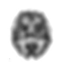
Single photon emission computed tomography
The single-photon emission computed tomography (short SPECT of English single photon emission computed tomography ) is a diagnostic method for producing cross-sectional images of living organisms and thus a variation of the emission computed . SPECT images show the distribution of a radiopharmaceutical in the body. Depending on the type of radiopharmaceutical, they are suitable for assessing the function of various organs.
Principle and implementation
Based on the principle of scintigraphy , the patient is given a radiopharmaceutical (a radionuclide or a substance labeled with a radionuclide) at the beginning of the examination , usually as an injection into a vein in the arm. The radionuclides used emit gamma radiation , which is detected with gamma cameras. One or more such cameras rotate around the body and detect the emitted radiation from different spatial directions. From these planar recordings (so-called projections), by means of inverse radon transformation, conclusions can be drawn about the distribution of the radiopharmaceutical inside the body. B. represent as sectional images through the body. In contrast to "static" SPECT examinations, in which only the distribution of the radiopharmaceutical is determined at a point in time, there are also so-called "dynamic" examinations, whereby one can assess the by repeated measurements at intervals of minutes, hours or days change in the radioactivity distribution over time (e.g. with 133 Xe ). SPECT is frequently used in the context of cardiology , whereby the measured decays are recorded in relation to the heartbeat (measured e.g. by an additional EKG ). The latter process is called gated SPECT , because the data is sorted into different gates or bins .
Further areas of application
- Myocard-SPECT to examine the blood flow (and restricted vitality) of the heart muscle tissue ( myocardial scintigraphy ). The radiopharmaceutical used is usually the technetium isotope 99m Tc in tetrofosmin or in MIBI (methoxyisobutyl isonitrile).
- Bone SPECT for the localization of regions with altered bone metabolism in the skeletal scintigraphy .
- Brain function SPECT: FP-CIT- (abbreviation for 123 I-N-ω-fluoropropyl-2β-carbomethoxy-3β- (4-iodophenyl) nortropane) and IBZM- (abbreviation for 123 I-iodobenzamide) SPECT for the diagnosis and differentiation of Parkinson’s syndromes and other degenerative brain diseases
- Epilepsy SPECT, see Brain Perfusion Scintigraphy
- Octreotide -SPECT in the context of somatostatin receptor scintigraphy in neuroendocrine tumors
- 123 I-Metaiodbenzylguanidin-scintigraphy ( MIBG-scintigraphy ) for adrenergic tumors z. B. the adrenal medulla, so-called. Phaeochromocytoma
Comparison and combination with other methods
Like positron emission tomography (PET), SPECT is one of the functional imaging processes : the images generated primarily provide information about metabolic processes in the body being examined. The morphology of the body, on the other hand, can only be roughly assessed, as this is not or only partially contained in the metabolic information shown and the resolution is also inferior to other methods. X-ray computed tomography (CT) is more suitable for showing the morphology.
Newer device systems such as SPECT / CT allow the combination of the advantages of morphological and functional imaging on one camera and data evaluation on the same computer system. The resulting so-called fusion images enable the precise assignment of functional abnormalities to the anatomical structures. This method is of particular importance when assessing various types of cancer and examining their progress.
Compared to PET, SPECT is less complex and cheaper because, on the one hand, no short-lived radionuclides are used, which have to be produced in close proximity to the scanner, and, on the other hand, the scanners are much cheaper (less electronics). Nowadays, however, the areas of application of the two processes are flowing into one another. The rapidly disintegrating radionuclides commonly used in PET are now also used in SPECT. The main disadvantages are the lower spatial resolution compared to PET and the lower sensitivity of the cameras. The reason lies in the camera principle, in which the directional information of the radiation is obtained by means of collimators , which in fact act like filters and keep almost all radiation away from the camera, except that which comes from a precisely defined direction. This considerably reduces the imaging efficiency related to the necessary use of the radionuclide compared to PET.
In nuclear medical diagnostics with SPECT spotlight γ-recourse (usually alone on 99m Tc ), since other types of radiation (α- and β - radiation) have a very short range in tissue, are measured at or outside of the body to be able to. These types of radiation are used in nuclear medicine therapy . β + emitters are used in PET, but there the photon emission (γ radiation) is used as a secondary effect ( annihilation radiation) ( triggered by the primary positron or β + particles ).
Web links
Individual evidence
- ^ MD Cerqueira, AF Jacobson: Assessment of myocardial viability with SPECT and PET imaging . In: American Journal of Roentgenology . tape 153 , no. 3 , 1989, pp. 477-483 ( PDF [accessed September 7, 2011]).
- ↑ W. Reiche, M. Grundmann, G. Huber: The dopamine (D2) receptor SPECT with 123I-iodobenzamide (IBZM) in the diagnosis of Parkinson's syndrome . In: The Radiologist . tape 35 , no. 11 , 1995, pp. 838-843 ( abstract ).
- ^ Gregor K Wenning, Eveline Donnemiller, Roberta Granata, Georg Riccabona, Werner Poewe: 123I ‐ β ‐ CIT and 123I ‐ IBZM ‐ SPECT scanning in levodopa ‐ naive Parkinson's disease . In: Movement Disorders . tape 13 , no. 3 , 1998, p. 438-445 , doi : 10.1002 / mds.870130311 , PMID 9613734 .
- ↑ Wim Van Paesschen, Patrick Dupont, Stefan Sunaert, Karolien Goffin, Koen Van Laere: The use of SPECT and PET in routine clinical practice in epilepsy . In: Current Opinion in Neurology . tape 20 , 2007, p. 194-202 , doi : 10.1097 / WCO.0b013e328042baf6 , PMID 17351491 .
- ^ Kjell Oberg: Molecular imaging in diagnosis of neuroendocrine tumors . In: The Lancet Oncology . tape 7 , no. 10 , 2006, p. 790-792 , doi : 10.1016 / S1470-2045 (06) 70874-9 , PMID 17012038 .
- ↑ Anders Sundin, Ulrike Garske, Håkan Örlefors: Nuclear imaging of neuroendocrine tumors . In: Best Practice & Research Clinical Endocrinology & Metabolism . tape 21 , no. 1 , 2007, p. 69-85 , doi : 10.1016 / j.beem.2006.12.003 .
- ↑ Vittoria Rufini, Maria Lucia Calcagni, Richard P. Baum: Imaging of Neuroendocrine tumor . In: Seminars in Nuclear Medicine . tape 36 , no. 3 , 2006, p. 228-247 , doi : 10.1053 / j.semnuclmed.2006.03.007 .
- ↑ Christoph Matthias Schmied: 123 I-Metaiodobenzylguanidin-Scintigraphy and magnetic resonance tomography in the detection of lesions in neuroblastomas in children and the importance of a combined diagnosis of both methods , dissertation, LMU Munich, 2005 ( PDF ).
