Scintigraphy
The scintigraphy or scintigraphy (from latin scintillating "spark" and ancient Greek γράφειν "drawing, describe") is an imaging technique of nuclear medicine functional and diagnostic localization. The image created after the administration of a radiopharmaceutical with organ-specific distribution is called a scintigram .
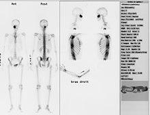
For a scintigram, radioactively marked substances (radiopharmaceuticals) are introduced into the body, which accumulate in the target organ to be examined and are then made visible with a gamma camera , which measures the emitted radiation. The method is not only suitable for localization diagnostics, for example of inflammation centers in the skeleton ( skeletal scintigraphy ), but also for the detection and display of an increased metabolism. Since the chronological sequence of the uptake and excretion of the radiating substance can be recorded, information about the function of organs can be obtained, for example, in kidney function scintigraphy or cardiac scintigraphy . The radiation exposure in these examinations is usually lower than in the comparable X-ray examinations . In Germany around 60,000 scintigraphies are performed every week.
principle

Imaging is based on the administration of radiopharmaceuticals; H. Substances that are radioactively marked. The substances used are those that accumulate particularly well in the tissue to be examined. One speaks of a tracer (indicator).
Radiopharmaceuticals that emit gamma rays are used. The radionuclides accumulate, depending on their chemical and biological properties, in certain body organs (e.g. thyroid , heart , liver , kidney , lungs , bones ). In skeletal scintigraphy , for example, bisphosphonates are used, which are incorporated into the bone substance as a result of metabolism. As radioactive marker is usually used technetium - isotope 99 m Tc.
With the help of a scanner or a gamma camera , the emitted radiation can be determined (detected) and transformed into a color-visualized image. A scintillation crystal is used for detection , which generates flashes of light when the gamma quanta hit. "Szinti" comes from the Latin scintillare and means "to flash, sparkle"; hence the name "scintigraphy". The flashes of light from the crystals are converted into an electronic signal and displayed as image points in degrees of blackening according to their frequency. The organs being examined can be displayed either flat (planar) or using SPECT . Several recordings of the same body region are made from different angles and a three-dimensional model is calculated from the data obtained, which enables sectional images like in a computer tomography .
application
Scintigraphy is used, for example, in tumor diagnostics. The radioactively marked tracer will preferentially accumulate in tissue that has an increased metabolism and is therefore more vascularized (blood supply) (“ hot spot ”). This is typical for tumor tissue. In the scintigram these tissue parts appear darker or stronger / differently colored. In the case of questions relating to the skeletal system, a comprehensive overview can be obtained very quickly with this procedure, and unclear findings can be specified more precisely. If, for example, a loosened endoprosthesis is suspected , the scintigraphy will reliably prove this or rule it out. The state of activity and the distribution pattern of a rheumatic disease can be estimated, the question of skeletal metastases can be answered.
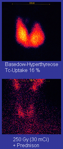
It is also possible to use this method to diagnose an increased or decreased metabolism of non-tumor tissue, for example with a thyroid examination. The illustration on the right shows an example.
Another possible application is in paediatrics and adolescent medicine: If there is a suspicion of abuse in children, especially infants ( Battered Child Syndrome - frequent clinical indication "falling from the changing table"), a scintigraphy can determine increased bone metabolism processes occur as a repair measure of the bone. It is thus possible to draw conclusions about the use of external violence. The bones do not have to be broken for this, even slight bruises can be detected with the help of scintigraphy.
The time span for the examinations is - depending on the underlying physiological processes - sometimes several hours; in skeleton scintigraphy z. B. three to four hours can be set from the administration of the radiopharmaceutical to the completion of the recordings. For the recording itself, the patient lies quietly under the gamma camera for 10 to 30 minutes, depending on the question and the device . The radiation exposure varies depending on the examination and is, for example, for a thyroid scintigraphy at the level of a simple X-ray (about 0.5 mSv ), for most examinations it is below that for a more extensive computed tomography (about 5 to 20 mSv), and in individual cases about that. The indication for a nuclear medicine examination is to be made strictly in children and adolescents, in pregnant women there is usually no indication. Furthermore, it should be borne in mind that the technology used is based on the fact that the radiation leaves the body. Depending on the radionuclide used, too close contact with pregnant women, children and adolescents should be avoided in the first 24 to 48 hours after the examination.
Similar procedures
See also
literature
- Cornelius Borck: Scintigraphy. In: Werner E. Gerabek , Bernhard D. Haage, Gundolf Keil , Wolfgang Wegner (eds.): Enzyklopädie Medizingeschichte. De Gruyter, Berlin / New York 2005, ISBN 3-11-015714-4 , p. 1375.
Web links
- Scintigraphy: images produced by radioactive substances , Cancer Information Service of the German Cancer Research Center (DKFZ), Heidelberg, March 24, 2010; Retrieved September 4, 2014.
- Nuclear Medicine Information - Nuclear Medicine Topic Collection
Individual evidence
- ↑ Der Kleine Stowasser , Munich 1971
- ↑ Michael Feld: Acute Bottlenecks in Nuclear Medicine . In: Dtsch Arztebl , 2008, 105 (37), pp. A-1874 / B-1614 / C-1578, current: Akut


