Pterygopalatine fossa
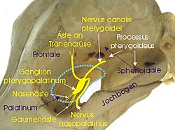
Pterygopalatine fossa (outlined in blue) and pterygopalatine ganglion in dogs
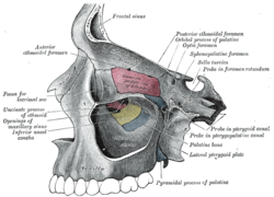
The pterygopalatine fossa (winged palatine fossa ) is a bony-limited pit (Latin fossa ) on the skull .
Limits
It has the following limits:
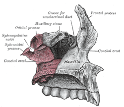
- Forward (anterior): Maxilla (Facies infratemporalis)
- backwards (posterior) and upwards (cranial): Os sphenoidale (Processus pterygoideus)
- to the middle (medial): Os palatinum (perpendicular plate)
- downwards (caudal): it narrows to the palatine canal major
- Lateral: pterygomaxillary fissure (passage of the maxillary artery, going from the infratemporal fossa to the pterygopalatine fossa)
links
The pterygopalatine fossa has connections to other parts of the skull:
- to the front (anterior) - via the inferior orbital fissure - to the orbit
- downwards (inferior) - over the palatine canal - to the oral cavity and the canali palatini minoris
- to the outside (lateral) - via the pterygomaxillary fissure - to the infratemporal fossa
- to the inside (medial) - via the perpendicular plate of the palatine bone with the sphenopalatine foramen - to the nasal cavity
- to the rear (posterior) - over the foramen rotundum - to the fossa cranii media
- backwards (posterior) - via the palatovaginal canal (pharyngealis) - to the nasal cavity ( nasopharynx )
- to the rear (posterior) - over the pterygoid canal (Vidi nerve) - fossa cranii media and foramen lacerum
content
The following anatomical structures are found in the pterygopalatine fossa:
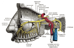
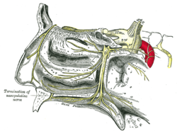
- Pterygopalatine ganglion (with connection to the maxillary nerve )
- the third section of the maxillary artery (pars pterygopalatina)
- Nerve in the pterygoid canal (N. petrosus major (parasympathetic) and N. petrosus profundus (sympathetic) = N. canalis pterygoideus)
