Juxtaoral organ
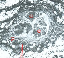
The juxtaoral organ ( Latin for “lying next to the oral cavity”, synonym: Chievitz organ, after Johan Henrik Chievitz, Danish anatomist (1850–1901), who first described this ramus mandibularis ductus parotidei in 1885) is a small elongated (10– 12 mm long, 2–3 mm thick) organ in the cheek, consisting of a bud-rich epithelial parenchyma , which is embedded in a connective tissue rich in nerves and cells and surrounded by a tight perineurium-like envelope.
Formerly known as a structure that only occurs during embryonic development (Chievitz's organ), it was detected by Zenker in 1953 as a regularly occurring organ in adults. Taking into account its localization in humans and animals, Zenker and Salzer gave it the name juxtaoral organ . This designation was accepted by the International Anatomical Nomenclature Commission and included in the Nomina anatomica .
topography
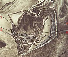
The juxtaoral organ lies in the deep lateral region of the face between the musculus buccinator and the temporalis tendon, under the fascia buccotemporalis near the point of passage of the parotid duct through the cheek muscle. The organ is innervated by the buccal nerve.

Feinbau
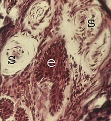
The histological picture shows numerous epithelial buds, embedded in connective tissue rich in cells and nerves. Sensory nerve cells end in many places on the epithelium. But there are also many specialized nerve end bodies .
development

In 11 to 15 mm human embryos, the development of the juxtaoral organ begins as a sprouting of an epithelial ridge (1) from the oral cavity epithelium of a side pocket ( sulcus buccalis ) of the embryonic oral cavity. As a result, the organ system together with the nerves are shifted away from the oral mucosa to the above-mentioned point in the deep facial region.
function
So far only morphological indications are available. A receptor function can be assumed because the organ is innervated by sensory trigeminal nerve branches and contains a large number of receptor nerve endings. It may perceive dynamic changes that occur in the act of chewing and swallowing.
Experimental studies on the rat suggest that the buccal nerve could exert a trophic influence on the organ. After removal of the pituitary gland in rats, considerable atrophy of the juxtaoral organ was observed (Salzer and Zenker).
Clinical significance
The organ can be used as a starting point for tumors.
The possibility of a misinterpretation of the organ in the biopsy as a carcinomatous structure has already occurred and resulted in a major operation. With regard to differential diagnoses, pathologists and oral surgeons are confronted with the fact that there are other epithelial structures in the immediate vicinity of the juxtaoral organ, such as salivary glands of the cheeks and epithelial nests that have formed from epithelial remnants of embryonic development.
further reading
- W. Zenker: Juxtaoral Organ, Morphology and Clinical aspects . Urban & Schwarzenberg, Munich 1982, ISBN 3-541-72221-5 .


