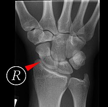
Scaphoid fracture
| Classification according to ICD-10 | |
|---|---|
| S62.0 | Fracture of the scaphoid of the hand |
| S92.2 | Fracture of one or more other tarsal bones naviculare pedis |
| ICD-10 online (WHO version 2019) | |
The scaphoid fracture or scaphoid fracture is a fracture (fracture) of a scaphoid bone - either the scaphoid of the wrist or the navicular of the tarsal. The scaphoid is shortened eingedeutscht also scaphoid called - is therefore also from a scaphoid fracture spoken. In the following, only the scaphoid fracture of the hand is shown.
Mechanism of Accident and Epidemiology
Injury to the navicular bone accounts for three quarters of all wrist fractures. It is usually caused by direct force, for example by falling on the backwards overstretched (dorsiflexed) hand with an impact on its spoke-side (radial) half. The exact height of the fracture depends on the strength of the hyperextension and lateral movement on the side of the spoke (radial abduction) of the hand at the moment of impact.
The injury is most common in sports, around 90% of patients are men, and the most common age group is those between 20 and 30 years of age.
Diagnosis
On examination, there is often pain on pressure, sometimes with swelling, in the area of the radial foveola , also known as a tobacco box. There, pain often arises when the examiner passive movement of the carpal wrist in pronation and elbow-sided lateral movement (to the ulnar) as well as pronation and lateral lateral movement (to the radial). Usually the mobility of the thumb is also reduced and the attempt to bring the thumb and index finger into opposition, as with a point grip, is painful or even impossible. Pain can also be triggered by axial compression of the thumb, as well as by pressure on the tuberculum ossis scaphoidei in the area of the distal crease of the hand with the wrist extended and held towards the spoke. In a clinical study, pain on pressure over the scaphoid tubercle and pain in the tobacco box with ulnar-sided movement of the wrist were shown to be the most sensitive diagnostic tests.
Nevertheless, the clinical signs have a low specificity overall , and X-rays do not initially show a fracture line in up to 40% of cases. In X-ray diagnostics, four images of the wrist including two oblique images ("scaphoid quartet") are made if there is a corresponding suspicion. Alternatively, in addition to the two standard exposures of the wrist, a single exposure can be made by Stecher, in which the hand is clenched into a fist and abducted in the ulnar direction. If a break cannot be determined with certainty despite clinical suspicion, computed tomography is usually carried out to confirm the diagnosis , or - especially in children for whom radiation protection plays an even greater role - magnetic resonance imaging .
treatment
Healing a scaphoid fracture in the hand is difficult, depending on the exact location of the break, since the blood supply is mostly remote from the body, and it can take eight to twelve weeks. During this time, the wrist with the base of the thumb is immobilized in a cast or a splint up to the forearm .
A good alternative to plaster treatment is to screw the navicular bone with a special cannulated screw with two threads of different pitches. This shape of the screw compresses the fracture fragments. The most commonly used screws of this type are the Herbert screw, named after Timothy James Herbert , and the Bold screw . The osteosynthesis is usually done through a small incision on the flexor side of the wrist. The advantage of the minimally invasive operation is the stable, secure treatment of the fracture and the considerably shorter post-treatment time due to the operation.
Complications
Scaphoid pseudarthrosis is a complication in the treatment of scaphoid fractures if the fracture does not heal .
Childish scaphoid fracture (hand)
A scaphoid fracture is very rare in childhood, accounting for only 0.34% of all child fractures and only 3% of all hand fractures. Earlier and especially before the age of ten, fractures of the distal pole, which first ossified, occur, which have a very good prognosis. Increasing sporting activity, which is increasingly similar to adult sports, has shown a different distribution pattern in children from 12 years of age in the last twenty years, which is more similar to that of scaphoid fractures in adults.
In a large case series from Boston with 351 childhood scaphoid fractures between 1995 and 2010, the average age was 14.6 years, the youngest child was seven years old. The fracture location was distal in 23%, central in 71% and at the proximal end of the navicular bone in 6% . The non-classical fractures in the middle and proximally occurred significantly more frequently in boys, in high-energy injuries, after growth plates had been closed and in the case of increased body mass index .
The therapy usually consisted of a three-month plaster immobilization. Delayed fracture healing was more common in delayed diagnosed fractures, displaced fractures, proximal fractures, and fractures with accompanying osteonecrosis . The plaster of paris immobilization of fresh fractures showed fracture healing in 90%, while the direct surgical stabilization of fresh fractures showed healing in 98%, but without the subsequent immobilization being shorter than in the non-operative group.
If the fracture does not heal, surgical intervention is indicated, which led to healing in 96%. As a rule, a cannulated screw is then passed through the two fragments, which presses the fragments together and secures them in a stable manner. Healing was delayed in children with still open growth plates, displaced ("displaced") fractures, proximal fractures, when using a bone chip, and depending on the type of screw used.
Almost a third of the children in the overview study showed a chronic delayed diagnosis of a hernia at the first presentation. The study suggests surgical stabilization directly because of the significantly lower probability of healing in the cast (only 23%).
literature
- S3 guideline for scaphoid fracture of the German Society for Trauma Surgery (DGU), the German Society for Orthopedics and Orthopedic Surgery (DGOOC) and the German Society for Hand Surgery (DGH). In: AWMF online (as of 2015)
- H. Krimmer et al. a .: Scaphoid fractures - diagnosis, classification and therapy. In: The trauma surgeon. 103, 2000, pp. 812-819. doi: 10.1007 / s001130050626 , ISSN 0177-5537
Web links
Individual evidence
- ↑ Bernhard Weigel, Michael Nerlich: Praxisbuch Unfallchirurgie. Springer-Verlag, Berlin 2005, ISBN 3-540-41115-1 , p. 375f.
- ↑ AD Duckworth, GA Buijze, M. Moran, A. Gray, CM Court-Brown, D. Ring, MM McQueen: Predictors of fracture Following Suspected injury to the scaphoid. In: The Journal of bone and joint surgery. Volume 94, Number 7, July 2012, pp. 961-968, doi : 10.1302 / 0301-620X.94B7.28704 , PMID 22733954 .
- ↑ a b c J. J. Gholson, DS Bae, D. Zurakowski, PM Waters: Scaphoid fractures in children and adolescents: contemporary injury patterns and factors influencing time to union. In: The Journal of bone and joint surgery. American volume. Volume 93, Number 13, July 2011, pp. 1210-1219, doi : 10.2106 / JBJS.J.01729 , PMID 21776574 .
