wrist

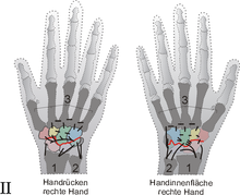
A – H = carpal bones
2 ulna ( ulna )
3 metacarpal bones ( ossa metacarpalia )

A navicular ( scaphoid )
B lunate ( lunate )
C triangle leg ( triquetrum )
D pisiform ( pisiform bone )
E Trapezium ( trapezium )
F trapezoid bone ( trapezoid )
G capitate bone ( Os capitatum )
H hook bone ( os hamatum )
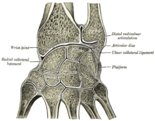
The wrist is the joints of several sub-composite joint ( articulatio composita ) at the hand of mammals . In the quadruped animals it is also called the pastern joint . In addition to the finger joints, it belongs to the joints of the hand ( lat. Articulationes manus ).
In humans, the joint between the spoke and the carpal bones ( articulatio radiocarpalis , " proximal wrist") and the joint between the two rows of the carpal bones ( articulatio mediocarpalis , " distal wrist") are referred to as the wrist. In a broader sense, the other joints of the wrist are also classified under this term, which, as so-called tight or loose joints ( amphiarthroses ), support the function of the two main joints , but only show a small range of motion. In most animals there is also a connection between the ulna and the carpal bones ( articulatio ulnocarpea ). In addition, the tight joints between the individual carpal bones ( Articulationes intercarpales ) and between the carpal and metacarpal bones ( Articulationes carpometacarpales ) are generally counted as part of the forefoot joint ( Articulatio carpi ).
All partial joints together act as a functional unit and allow diffraction ( flexion ) in the direction palm ( palmar flexion ), stretching ( extension ) direction of hand back ( dorsal ) and Abspreizbewegungen ( abduction ) direction thumb ( radial deviation ) and the direction of the little finger ( ulnar deviation ).
Proximal carpal joint
In humans, the proximal carpal joint, which is closer to the center of the body, denotes the articulated connection between the distal end of the spoke ( facies articularis carpi radialis ) and three of the proximal carpal bones ( ossa carpalia ), the scaphoid bone ( os scaphoideum ), the lunate ( lunate ) and the triangle leg ( triquetrum ). In addition, the inter -joint disc ( Discus triangularis ) of the distal radial-ulnar joint is involved in the formation of the joint, which mediates between the carpal bones and the ulna. However, in most vertebrates, both ulnocarpea ( ulnocarpea ) and radiocarpea ( articulatio radiocarpea ) are connected to the carpal bones.
A thin, slack joint capsule is attached to the cartilage margins of the bones and to the inter-joint disc, which is reinforced by numerous radiating ligaments . The joint space is unbranched and sometimes contains fat folds ( plicae synoviales ).
From a functional point of view, the proximal wrist of humans is an ellipsoid or egg joint ( Articulatio ellipsoidea ), which has two degrees of freedom :
- Flexion ( palmar flexion , towards the palm of the hand) up to around 80 ° and extension ( dorsiflexion , towards the back of the hand) up to around 70 °
- Spreading movements ( abduction or adduction ) towards the spoke ( radial abduction , actually more precisely radial adduction , i.e. towards the thumb side) up to approx. 20 ° or towards the ulnar ( ulnar abduction , towards the little finger side) up to approx. 40 °.
Distal carpal joint
The carpal joint ( articulatio mediocarpalis ) further away from the middle of the body - rarely also referred to as the middle carpal joint, in animals representing the middle level of the forefront joint - shows an approximately S-shaped joint gap between the proximal and distal row of the carpal bones. It is made up of the individual joints between two adjacent bones and works functionally with the articulated connections of the carpal bones of a row with one another ( articulationes intercarpales ) as a unit. The joint capsule is tight on the palm of the hand, but slack on the back of the hand and forms numerous fat folds.
The wrist, which is further away from the center of the body, is a toothed hinge joint ( articulatio ginglymus ). The schematic course is shown in Figure II as a red line. Its range of motion is restricted by its curved shape as well as ligaments and joint capsules. It works together with the proximal wrist as a functional unit.
Intercarpal joints
The intercarpal joints ( Articulationes intercarpales ) denote the articulated connections between the carpal bones of a row.
The intercarpal joints are - like the carpometacarpal joints - so-called wobbly joints ( amphiarthroses ), which are so stiffened by numerous ligaments that they can hardly be moved. The joint capsule is slack from the center of the body and taut further away from the center of the body. These are so-called secondary joints, which increase the mobility of the carpal bones among each other and thus increase the mobility of the adjacent main joints ( articulatio radiocarpalis and articulatio mediocarpalis ).
The intercarpal between the pisiform bone ( pisiform ) and the triangle leg ( triquetrum ), the so-called pisiform joint ( articulatio ossis pisiformis or Articulatio ossis carpi accessorii ) is an independent joint with an independent joint capsule and independent joint space.
Metacarpals
Carpometacarpal joints
The carpometacarpal joints, the connection of the distal carpal bones with the second to fifth metacarpal bones ( Articulationes carpometacarpales II – V ) are not assigned directly to the wrist in humans, but always to the forefoot joint in animals. Like the intercarpal joints, they are so-called wobbly joints, which are so stiffened by numerous ligaments that they can hardly be moved. These are secondary joints that increase the mobility between the carpal bones and the metacarpal bones and thus increase the mobility of the neighboring main joints ( articulatio radiocarpalis and articulatio mediocarpalis ).
The thumb saddle joint ( Articulatio carpometacarpalis pollicis or Articulatio carpometacarpalis I ) is an exception to the five carpometacarpal joints, as, in contrast to them, it does not form a wobbly joint, but a real and thus freely movable joint. It denotes the articulated connection between the large polygonal bone ( Os trapezium ) and the metacarpal bone of the first finger , i.e. the thumb ( Os metacarpale I or Os metacarpale pollicis ). It is a saddle joint ( articulatio sellaris ) and is mainly required for the opposition of the thumb to the other fingers and is therefore of central importance for human grasping.
Intermetacarpal joints
The intermetacarpal joints ( Articulationes intermetacarpales ) are the articulated connections between the metacarpal bones of a row. Like the intercarpal joints and the carpometacarpal joints, they are amphiarthroses, which are so stiffened by numerous ligaments that they can hardly be moved. They increase the mobility of the metacarpal bones among each other and thus the mobility of the neighboring main joints ( articulatio radiocarpalis and articulatio mediocarpalis ).
Tapes
The individual joints of the wrist are supported by a multitude of ligaments that strengthen the joint capsule and guide movements. For a schematic overview, see schematic drawing II.
- Mediate between the forearm bones and the proximal carpal bones
- Sidebands
- lateral elbow ligament ( ligamentum collaterale carpi ulnare ) and
- lateral radial ligament ( ligamentum collaterale carpi radiale ),
- Spoke hand bands
- Palmar radiocarpal ligament and
- Dorsal radiocarpal ligament ,
- Elbow -hand ligament ( palmar ulnocarpal ligament ).
- Sidebands
- Mediate between adjacent carpal bones
- Internal bands
- Ligamentum carpi radiatum : It describes a complex of ligaments ( ligamentum radiocarpale palmar , ligamentum intercarpale palmar and ligamentum carpometacarpale ) on the palm of the hand, which extend from the cusp of the head bone ( os capitatum ) and house all flexor tendons with the median nerve ( nervus medianus ),
- Pisohamate ligament and
- Flat ligaments ( ligamenta intercarpalia )
- Ligamenta intercarpalia palmaria ,
- Ligamenta intercarpalia dorsalia and
- Intercarpal ligament .
- Internal bands
- Between the carpal and metacarpal bones ( Ligamenta carpometacarpalia )
- Pisometacarpal ligament ,
- Palmar carpometacarpal ligament and
- Ligamentum carpometacarpal dorsal .
- The carpal ligament ( retinaculum flexorum or ligamentum carpi transversum ) spans across the carpal bones, which extends from the hook of the hook bone ( Hamulus ossis hamati ) and the pea bone ( Os pisiforme ) to the cusp of the large polygonal bone ( tuberculum ossis trapezii ) and the scaphoid bone ( Tuberculum ossis scaphoidei ) runs. It thus closes a bony connective tissue (osteofibrous) canal, the carpal tunnel ( Canalis carpi ).
- On the back of the hand, the extensor tendons run in the tendon sheath (also called tendon fans).
development
At first glance, the upper extremities of humans seem to have little in common with the pectoral fins of fish , but the basic structure is definitely comparable.
As can be seen in Figure III, ulna (1) and radius (2) have remained almost unchanged in the course of development. Likewise, the metacarpal bone ( metacarpal ) (3). There has been a significant reduction in the carpal bones. The proximal row of the carpal bones closer to the center of the body is punctured. The centralia no longer present in humans brown. The carpal bones further away from the center of the body (distal row) are hatched.
In birds, carpal bone reduction is most advanced. From the proximal row, only the os carpi radiale and ulnare are present, while the distal row has grown together with the metacarpal bones to form the carpometacarpus (→ bird skeleton ). For this reason, there are only two levels of joints in the wrist of birds - between the forearm and carpal bones and the carpal bones and carpometacarpus.
Diseases
One of the most common diseases of the wrists is carpal tunnel syndrome (CTS). The middle arm nerve ( nervus medianus ) is usually damaged by pressure. The cause can be previous broken bones near the wrist ( fractures ), rheumatic diseases or overuse ( computer mouse , bicycle handlebars, etc.). However, there is often no determinable cause. Damage to the ulnar nerve in the area of the wrist ( Loge de Guyon syndrome ) is much less common.
The most common damage caused by injuries ( trauma ) is a fracture of the spoke near the joint ( distal radius fracture ) and a fracture of the scaphoid bone . In principle, all bones of the wrist can break or get out of their structure if ligaments tear . Other diseases are tendinitis , repetitive strain injury syndrome and a ganglion . An arthrosis of thumb saddle joint is quite common and is used as rhizarthrosis referred.
In domestic dog puppies , the developmental disorder can be the carpal laxation syndrome , which is characterized by over- or under-stretching.
Investigation options
In addition to computed tomography (CT) and magnetic resonance imaging (MRT), a modern form of diagnosis is scintigraphy and wrist mirroring ( arthroscopy ), which can take place under local or arm (brachial plexus) anesthesia. The latter should only be carried out at centers where appropriate therapy can be carried out immediately , otherwise a second operation under anesthesia is often necessary with all the risks.
See also
Individual evidence
- ^ Roche Medical Lexicon
- ^ Franz-Viktor Salomon: Bone Connections. In: Franz-Viktor Salomon et al. (Hrsg.): Anatomie für die Tiermedizin. Enke-Verlag, Stuttgart 2004, ISBN 3-8304-1007-7 , pp. 110-147.
- ^ Alfred Sherwood Romer, Thomas S. Parsons: Comparative Anatomy of Vertebrates. 5th edition. Paul Parey Verlag, Hamburg 1983, ISBN 3-490-21718-7 .
- ^ Franz-Viktor Salomon: Textbook of poultry anatomy . Gustav Fischer Verlag, Jena 1993, ISBN 3-334-60403-9 .


