Triangular leg

A – H = carpal bones
2 ulna ( ulna )
3 metacarpal bones ( ossa metacarpalia )

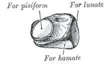
The approximately pyramid-shaped triangle leg ( lat. Triquetrum or carpal ulnar , fr. Os pyramidal ) is one of the eight carpal bones , is the proximal ( proximal ) series of these short bones on.
Its apex ( apex ossis triquetri ) is directed towards the middle ( medial ) and the base is on the side ( lateral ). This forms an articulated connection with the lunar bone ( os lunatum ). Form ( proximal ) the triangle leg is connected to the flexible disk ( articular ) and distally ( distally ) with the hook leg ( hamate ) in combination. On the palm of the hand ( palmar ) there is a small joint surface for the pea bone ( os pisiforme ).
Traumatology
Fractures of the cuneiform are rare, the triangle leg but after the scaphoid second most of all carpal bones affected. Often the fractures cannot be seen on standard X-rays of the hand, which is why the diagnosis is made using computed tomography if there is a suspicion . 90% of all fractures are dorsal avulsion or chipping fractures with a small, slightly dislocated bone fragment, triggered either by a ligament tear of the complex dorsal wrist ligament or by impacting the styloid process of the ulna and hookbone . Other forms of fracture include a palmar avulsion fracture and fractures that go through the body. While the avulsion or chipping fractures usually heal with a four to six week cast immobilization, it may seldom be necessary to surgically reduce the fractures and fix them with a screw osteosynthesis .
literature
- W. Platzer: Pocket Atlas of Anatomy, Volume 1 - Musculoskeletal System. Thieme Verlag, Stuttgart 2005, p. 126. ISBN 3-13-492009-3
- Christian Schuster: Chapter 7.2.3. - Other carpal fractures , pages 384–387 in chapter 7: Hand in: Bernhard Weigel, Michael Nerlich (eds.): Praxisbuch Unfallchirurgie Volume 1, Springer-Verlag Berlin 2005, ISBN 3-540-41115-1
