Triangular fibrocartilaginous complex
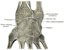
The triangular fibrocartilaginous complex ( Engl. : Triangular fibrocartilage complex , abbreviated TFCC ; lat. : Triangular fibrocartilage or articular ulnocarpalis ) in man is a triangular shaped, of fibrocartilage existing intermediate joint disk at the wrist , both the distal ends of ulna ( ulna ) and The spoke ( radius ) is firmly anchored to one another, as well as connecting them to the carpal bones ( ossa carpi ). In its function, the TFCC is comparable to the meniscus .
anatomy
The TFCC is a double-curved ( biconcave ) disc and is up to 5 mm thick at its edges. It is located on the wrist between the ulna with the joint space between it and the radius ( articulatio radioulnaris distalis ) and the proximal row of the carpal bones with the joint space between them and the radius ( articulatio radiocarpalis ). It is attached to the radius between the ulnar incisura and the articularis carpalis surface , and to the ulnar to the styloid process .
The TFCC is supplied with blood through dorsal and palmar , radioulnar branches of the ulnar artery and the anterior interosseous artery . The distribution of the supplying vascular branches ( microvascularization ) is comparable to the menisci on the knee: the edges ( periphery ; corresponds to 10–40% of the structure) are well supplied with blood, while the center and interior of the TFCC are barely reached.
The name "triangular fibro-cartilaginous complex" goes back to the surgeons AK Palmer and FW Werner, who first described the TFCC in 1981 as a homogeneous structure made up of:
- the articular disc,
- the dorsal and volar ligaments of the wrist,
- a meniscus-like structure,
- the ulnar collateral ligament of the wrist, as well
- the tendon sheath of the extensor carpi ulnaris muscle .
function
When the spoke moves in the wrist, the TFCC is taken along and rotates under the distal joint surface of the elbow. In addition, it is important as a joint connection (“intra-articular ligament ”) between ulna and radius - in the so-called distal radioulnar joint (DRU joint) - the stability of which is primarily ensured by the TFCC.
Clinical significance
In older people, degeneration causes the TFCC to fray. It is particularly affected by rheumatic diseases in the wrist, the progressive destruction of which also affects the distal radioulnar joint and the 6th extensor tendon compartment.
In young people, on the other hand, injuries can result in an acute rupture due to the fact that the TFCC is thinner in the middle and more vulnerable. There is pain in the ulnar wrist, especially when the hand is bent towards the ulnar. Swelling can develop over time, and some patients also notice a "cracking" sound when they move their hands.
examination
With the TFCC load test , twisting the wrist in maximum overstretching towards the back of the hand ( dorsiflexion ) can trigger pain on the side of the elbow (this is called a positive TFCC load test). By magnetic resonance imaging (MRI) can lesion with a sensitivity of 100% and a specificity are well represented by 90%. X-rays are usually inconspicuous and of little help. However, complaints due to an injury to the TFCC are relatively rare. Therefore, in the first place another, more likely cause should be ruled out.
therapy
Degenerative diseases of TFCC are usually treated conservatively with local anti-inflammatory drugs (e.g. ointments), cooling and possibly immobilization. In the case of advanced osteoarthritis or greater instability of the joint, the TFCC, including the ulnar wrist ligaments, can be tightened as part of the Sauvé-Kapandji operation ( joint stiffening of the distal radioulnar joint with simultaneous segmental resection of the ulna). If the symptoms persist, the TFCC can be surgically removed as a last resort . However, this method is not recommended because of possible chronic pain or subsequent joint instability . In young people, an acute tear can also be sutured.
literature
- A. Berger, R. Hierner: Plastic Surgery IV - Extremities , Volume 4. Springer Verlag, Berlin, 2008. ISBN 3540001441 .
- J. Duparc et al .: Surgical Techniques in Orthopedics and Traumatology - Wrist and Hand (corresponds to: Volume V). Lehmanns special edition, Urban & Fischer Verlag, Munich, 2005. ISBN 978-3-86541-284-3
- W. Graumann, D. Sasse, et al .: Compact textbook of the entire anatomy , Volume 2 ( musculoskeletal system ). Schattauer GmbH, 1st edition, 2004, ISBN 3794520629 .
- AK Martini: Orthopedic hand surgery: Manual for clinic and practice , 2nd edition. Steinkopff Verlag, Würzburg, 2008. ISBN 3798515255 .
- N. Wülker et al .: Pocket Textbook Orthopedics and Trauma Surgery , 1st edition. Thieme Verlag, Stuttgart, 2005. ISBN 3131299711 . P. 392 ff.
Individual evidence
- ↑ a b c Wülker, 2005, p. 412
- ↑ Gaumann / Sasse, 2004, p. 612
- ↑ cf. MS Bednar et al .: The microvasculature of the triangular fibrocartilage complex: Its clinical significance. J Hand Surg [Am]. 1991; 16: 1101-5
- ↑ a b cf. AK Palmer, FW Werner: The triangular fibrocartilage complex of the wrist; anatomy and function. J Hand Surg [Am]. 1981; 6 (2): 153-62.
- ↑ Wülker, 2005, p. 395
- ↑ Martini, 2008, p. 92
- ↑ Berger / Hierner, 2008, p. 66
- ↑ cf. HG Potter et al .: The utility of high-resolution magnetic resonance imaging in the evaluation of the triangular fibrocartilage complex of the wrist. In: J Bone Joint Surg Am . 1997; 79 (11): 1675-84.
- ↑ Martini, 2008, p. 53
