
roentgen


Röntgen (named after the physicist Wilhelm Conrad Röntgen ), also known as X-ray diagnostics , is a widely used imaging method in which a body is irradiated using an X-ray tube. The penetration of x-rays into the body is represented in images called x-rays , x-rays, or radiographs . Overall, the technical devices for imaging are referred to as X-ray apparatus or X- ray apparatus .
The images are visible on a fluorescent screen , for example . When fluoroscopy with an x-ray camera one is x-ray image intensifier needed. Suitable film material can also be used (radiography with X-ray film ). However, the state of the art is digital x-ray (digital radiography). Phosphor plates ( X-ray image plates ) or electronic sensors, for example CCDs, are used here . The medical procedures are detailed under Radiology .
history
On November 8, 1895, Wilhelm Conrad Röntgen discovered the invisible rays in Würzburg. He experimented with a nearly evacuated cathode ray tube made of glass. He covered it with cardboard, but the rays could penetrate it and showed an object randomly on the table on the fluorescent screen. On December 28th, he submitted his first written communication “About a new kind of rays” to the Physical-Medical Society in Würzburg, and on January 23rd, 1896, his discovery was first publicly demonstrated. He renounced a patent so that the X-ray machines could be used more quickly. In 1901, Röntgen received the first Nobel Prize in Physics for his discovery, which was used worldwide from 1896/1897. Based on Röntgen's discovery, Carl Heinrich Florenz Müller developed the first water-cooled anode together with doctors .
In the German Roentgen Museum in Roentgen's birth Remscheid - Lennep numerous historical X-ray machines are on display.
Use in medicine


principle
In medicine , X-rays are used to identify abnormalities in the body that, in connection with symptoms, signs and possibly other examinations, enable a diagnosis (X-ray diagnostics). The tissues of the human (or animal) body of different densities absorb the X-rays to different degrees, so that an image of the inside of the body is achieved ( shading , lightening and other X-ray signs ). The procedure is often used, for example, if a bone fracture is suspected : If the X-ray shows an interruption in the continuity of the bone, the suspicion is confirmed.
The conventional X-ray image shows an image of the three-dimensional object (e.g. an ankle joint - collapsed: ankle) on a two-dimensional surface. Therefore, many objects - such as extremities with questionable broken bones - are X-rayed from two directions (in technical jargon: "in 2 planes"). What is not yet noticeable from one perspective (or viewing direction) may do so from the other. Or if two bone parts of a fracture lie one behind the other in one direction, a displacement of the ends of the bone fracture (in technical jargon: "dislocation or dislocation") can only be shown on a second image from a different direction. For this purpose, standard recording techniques are available for almost all body parts that can be represented, so that the viewer does not have to “think his way around” every time. If the doctor arranges X-rays of an ankle joint in two planes, he can assume that he has a lateral (in technical jargon: "transversal") image showing the articular surfaces of the shin and talus (and a few others), as well as an image from the front rear (in technical jargon: ap = anterior - posterior) with easily assessable inner and outer ankles. If it is not yet clear, a slice image may be arranged in order to obtain sectional images instead of the simple "overview images".
Layered x-rays are called tomography . The more modern X-ray computed tomography (CT) differs from the “conventional tomography” ( X -ray tomography ). In this case, a computer calculates the sectional images from the electronic data that are generated from different directions during X-ray images. CT images have a much higher image quality.
Contrast media are often administered to the patient during or before the X-ray examination . Some structures that cannot normally be delimited can be highlighted in this way. In some cases, the function of an organ system can also be represented with a contrast agent, for example in urography . Depending on the question, there are various substances and dosage forms available.
In order to be able to clearly recognize the spatial position of broken bones or dislocated joints in particular, two to three images are usually made from a point in the body from different projection directions.
In addition to still images - at least since 2007 - X-ray videos can be filmed and displayed live on the screen when bone parts are reduced or adjusted in order to avoid opening the body with a scalpel incision and still obtain an informative picture of the changing position of the bones. The hands of the trauma surgeon working in the irradiated operating area are protected with lead-rubber gloves as far as possible.
Soft and hard radiation
Different "radiation qualities" are required for different areas of the body in order to produce tissues of different densities, such as B. to penetrate adipose tissue or bones. In X-ray diagnostics, one speaks of soft and hard radiation. The decisive factor is the voltage in kilovolts (kV) that is fed to the X-ray tube . Depending on the area of the body to be imaged or the desired image information, the tube voltage is selected between around 25 and 35 kV for mammography and around 38 and 120 kV for the other body regions.
The softer the radiation (low kV values), the greater the proportion of radiation absorbed by the tissue. This makes even the finest differences in tissue visible on the X-ray film. This is the case with mammography (X-ray examination of the female breast), but the radiation exposure of the irradiated tissue is relatively high. Hard radiation (over 100 kV) penetrates tissue and materials (plaster of paris and even lead aprons of smaller thickness) much more easily. Differences in contrast are greatly reduced, such as B. with lung exposures (120 kV), in which otherwise no assessment of the lung structure would be possible in the area of the ribs.
hazards
Not only X-rays lead to radiation exposure. Every year we are exposed to a natural radiation load of ~ 2.4 mSv / a, which is composed of cosmic radiation 0.3 mSv / a, earth radiation 0.5 mSv / a, natural radon inhalation 1.3 mSv / a and the absorption of natural radioactive sources Substances 0.3 mSv / a.
In addition, there is ~ 1.53 mSv / a civilization radiation exposure, of which from nuclear facilities <0.01 mSv / a, further caused by the use of radioactive substances and ionizing radiation in research, technology and the household <0.01 mSv / a, further caused by the case -out of nuclear weapons tests <0.01 mSv / a and finally the use of radioactive substances and ionizing radiation in medicine cause the majority of 1.5 mSv / a. The radiation exposure in medicine thus has a not inconsiderable share in the total radiation exposure of the population, but by far the largest share is accounted for by a few seriously ill patients.
The average radiation exposure from the Chernobyl reactor accident (April 26, 1986) in 1990 in Germany was 0.025 mSv. A 10-hour flight is equivalent to 0.1 mSv. The cosmic radiation exposure at 2000 m above sea level is 0.6 mSv higher. The regional difference in natural radiation within houses in Germany is 0.6 mSv.
The examples listed above are intended to serve as a benchmark for the following list of X-ray examinations. A one-time X-ray examination of the following type of examination results in an additional effective dose:
| X-ray examination | additional effective dose |
|---|---|
| Teeth / jaw | 0.02 mSv |
| skull | 0.2 mSv |
| Ribs | 3.0 mSv |
| Thorax (lungs) | 0.2 mSv |
| Abdomen | 0.3 mSv |
| Cervical spine | 2.0 mSv |
| Thoracic spine | 5.0 mSv |
| Lumbar spine | 0.4 mSv |
| pool | 0.1 mSv |
It usually takes years for radiation-induced cancer to develop. For leukemia (blood cancer), in this dose range, 15 years are assumed, for other forms of cancer it is 40 years.
Since the radiation doses used in X-ray diagnostics are potentially harmful for the patient and the user, special emphasis is placed on radiation protection in radiology . In Germany, in the event of an X-ray examination, the examining doctor offers patients to enter information such as the date and the irradiated body region in an X-ray passport or to have such a passport issued. The surgeon's safety is guaranteed by the fact that he has to press a key in an adjacent room, without which the X-ray apparatus cannot work. Keeping the release button pressed while observing the patient prevents the X-ray from being triggered in an uncontrolled manner or from being continued unintentionally if the surgeon faints.
Every year billions of images are taken around the world using radiation technology - around a third of these images in patients with acute myocardial infarction. Between 1980 and 2006 the annual dose increased by an estimated 700%.
Germany takes a top position when it comes to x-rays: around 1.3 x-rays and 2 mSv per inhabitant per year. Theoretically 1.5% of annual cancer cases can be traced back to this radiation exposure . In the opinion of the pediatric radiologist Christoph M. Heyer, doctors underestimate the radiation exposure from computed tomography: In 2003, these accounted for a good 6% of all X-ray examinations, but were responsible for more than 50% of medical radiation exposure .
Example: In the case of a coronary examination using computed tomography (CT), patients buy the increased sensitivity with an increased risk of cancer. American scientists calculated that one of 143 women examined by means of coronary CT in twenty-year- olds will develop cancer in the course of their life as a result of this angiography radiation, but only one of 686 men of the same age. CT angiography of the coronary arteries seems to increase the risk of cancer not insignificantly, especially in women and young people. When a patient comes to the clinic with an acute myocardial infarction , he is often given a radiation dose of 14.5 mSv in total, which corresponds to about 73 chest X-rays. The dose that a heart attack patient receives through this catheter examination corresponds to 3/4 of the statutory annual dose for workers in German nuclear power plants (20 mSv / a).
In a large-scale study, they analyzed the data of 64,074 patients who had been treated for an acute heart attack in teaching hospitals in the USA between 2006 and 2009 . A total of 276,651 examinations with ionizing radiation were carried out on this collective during this period. 83% of heart attack patients received chest x-rays, 77% catheter examinations. In the opinion of the speaker, necessary examinations that contain ionizing radiation should not be omitted - but one should be sure that these are appropriate.
Studies by US researchers have shown that frequent x-raying of the teeth triples the risk of benign brain tumors , and even quintuples in children under ten.
The conditions under which a doctor is liable for skin damage due to an X-ray examination is the subject of a decision by the Jena Higher Regional Court .
Contrast and contrast media
The absorption of X-rays depends on their energy level (achieved via different acceleration voltages) and increases with the number of atoms “in the way”, i.e. the thickness of the object and its atomic density (atoms / volume) as well as the atomic number (atomic number) ) Z and mass number A (atomic mass M as a guide) of the atoms of the material. Hollow organs (respiratory tract, lungs, stomach, intestines, bladder) or body cavities (abdominal cavity) can be represented by their content of air (gas), possibly inflated (with air, nitrous oxide, helium) by little absorption in the gas. On the other hand, bones are depicted as a shadow by the comparatively "heavy" calcium atom (Z = 20 / A = 40) if there is essentially water and hydrocarbons with oxygen as the heaviest atom (Z = 8 / M = 16). Dental and joint prostheses made of titanium (Z = 22 / M = 47) stand out from the bone due to their increased absorption. Those based on steel (iron: Z = 26 / M = approx. 56) are even stronger, as are the right / left marking rings made of NiRo steel or brass.
Historically, thorium (90/232) ( Thorotrast ) was used in angiography as an early or first contrast medium, but was banned around 1955 because of its radioactivity. Barium sulfate BaSO 4 (Ba: 56/137) in an aqueous suspension is used to map the gastrointestinal tract and the speed of the passage. Organic iodine compounds (I: 53/127) (iodine: 53/127) for angiography (intravenous or intraarterial) early Per-Abrodil = diethanolamine-3,5-diiodopyridone-4-acetic acid with acute side effects, later tolerated aromatic iodine derivatives.
Analog and digital X-ray
Digital X-rays are now considered the standard in imaging diagnostics . Digital X-ray has great advantages over the conventional analog process. The most important point is the reduction of radiation exposure .
Advantages of digital X-ray compared to analog X-ray:
- Reduction of radiation exposure
- The images are not overexposed or underexposed
- Recordings available immediately
- Post-processing on the computer
- Neither a darkroom nor a processor with consumables is required
- Reduction of the environmental impact
The storage of the digital X-ray images is standardized. This enables doctors to forward the files.
Picture gallery

Historical photo of a hand with a ring ( Röntgen , January 23, 1896)

X-ray machine from Siemens, photo 1950

Left broken upper arm
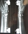
X-ray image of a comminuted radius fracture with plating and external fixator

A modern image of a left hand with 6 fingers ( polydactyly )
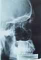
X-ray image of a male skull
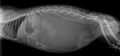
X-ray image of a dwarf rabbit
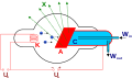
Schematic representation of an X-ray tube


Further areas of application in science
biology
In biological departments, such as zoology , attempts are made to answer a wide variety of questions with the help of X-ray-based representations. For example, the structure of the circulatory system in invertebrates and its position in the body can be examined better and faster than would be possible with conventional methods such as dissection under the microscope or histological sections.
Structural analysis
By measuring the diffraction of X-rays when passing through a substance sample, the crystal structure of substances can be clarified. Molecules can be visualized in this way. In the case of organic molecules such as DNA , RNA and proteins , the structure allows conclusions to be drawn about the function, which is why molecular biologists use X-ray structure analysis particularly often. The individual processes involved in this procedure are explained in the article Crystal structure analysis .
In addition to X-ray diffraction, X-ray absorption can also be measured. This is used in X-ray absorption spectroscopy as a method for structure elucidation. The method is not limited to crystalline samples, but it is only suitable for the elucidation of near structures. In the area of biological samples in particular, X-ray absorption spectroscopy is increasingly used for the targeted elucidation of active centers of enzymes.
Geology and mineralogy
The chemical analysis of rocks and minerals is possible with the help of X-ray fluorescence analysis. By irradiating with X-rays of approx. 50 kV, the chemical elements contained in a sample are excited to fluorescence radiation, the wavelength of which is characteristic of the element in question. The elements can be qualitatively determined by measuring the wavelength of this radiation. A quantitative analysis can also be carried out by measuring the intensity and comparing it with a standard sample of known composition. In contrast to wet chemical analysis methods, the method is non-destructive, i. i.e., the sample is unchanged after analysis and can be used for other purposes. However, a geological sample must be finely ground and pressed into a flat tablet (usually with a binder).
archeology
In archeology , for example, x-rays are used to x-ray mummies if their bandages are not to be destroyed. In addition, finds with a complex structure such as weapons, decorated ornaments or objects under lock and key in chests without opening can be examined.
Painting examination
Kurt Wehlte used X-ray technology for the first time to make the various layers of the image structure visible in paintings. He founded the X-ray image office for painting examination in Berlin .
Other technical applications
safety

At some checkpoints, X-ray technology is used in scanners to scan cavities or people in a time-saving but effective manner.
There are X-ray devices that can x-ray entire truck loads or containers, as well as mobile devices that are designed to x-ray an entire aircraft.
X-ray technology is also used to aid in the dismantling of bombs; this is for analysis .
material testing
Other applications can be found in X-ray materials testing . By X can be during the radiographic examination examine objects on cracks and voids in the interior. This is done with so-called X-ray refraction systems, usually with a loading mechanism to easily open the micro-cracks (crazes).
Quality control in food production
Large retail chains are increasingly demanding better detection of foreign bodies from food manufacturers in order to increase product quality. After the metal detector was the method of choice in recent years, X-ray systems are now being used more and more frequently. These X-ray systems consist on the one hand of the known X-ray system (tube / collimator and receiver) as well as of an advanced computer-aided image processing with control device. This means that the X-ray image of the respective food is examined for possible impurities (contamination) using special computer programs. If the X-ray image analysis shows that a food is contaminated, the connected control device is immediately informed that this food is to be controlled. It ends up in the trash can.
However, at the beginning of the use of such X-ray systems in the food industry, hurdles have to be overcome. The fear of exposure to possible radiation is often great and needs to be explained. Apart from X-ray systems that irradiate food in order to make it more durable, the X-ray examination with regard to possible contamination has absolutely no effect on the food itself. The X-ray has neither a preservation nor a destructive effect. What remains is the safety of the X-ray system for the user. Since X-rays in Germany require approval according to the regulation on protection against damage caused by X-rays , the hurdles for possible injuries are very high. Ultimately, the respective security depends on the operator himself and the purchased system. However, it should not be forgotten that medical X-rays and air travel (at normal altitude) temporarily entail far greater stress than is the case with an X-ray system for quality assurance. Anyone who is in the damp cellars of houses or in waterworks will usually get higher readings on the measuring device (dosimeter) than in front of the activated X-ray system. The radiation comes to us from the ground as well as from the stone walls and space and is also measured.
An X-ray system can detect metallic and non-metallic contamination, but not all. At the present time (2005), X-rays are the only way to identify as many and different small contaminations as possible in food. However, the assumption that the product is 100% contamination-free after the examination is incorrect. It is certain that in the coming years the detection capacity can be increased even further by means of better technology. But you will never be able to find everything. This is primarily due to the fact that the closer the "X-ray effects" of contamination and the actual product are to one another, the more difficult it is for the image processing system to differentiate between the two. The so-called Hounsfield scale lists the X-ray effects of a wide variety of materials. The closer the respective materials are in this list, the more difficult it is for an X-ray detector to distinguish them (example: meat and fat). If, on the other hand, the difference is great, such as B. between a piece of cheese (packed or unpackaged) and a small stone or piece of iron or aluminum, it is particularly easy for the X-ray detector to identify and sort out the impurities in the cheese.
See also
- Flat panel detector for X-rays
- History of radiation protection
- Gustav Peter Bucky (radiologist)
- Crystallography
- Pedoscope (historical use of X-ray machines in shoe shops)
- PIXE (particle-induced X-ray emission or proton-induced X-ray emission)
- radiology
- X-ray film
- " X-ray portrait "
- Radiation exposure
- Radiation sickness
literature
- EC Petri: The X-Ray Film. Properties and processing. Photo cinema, Halle 1960.
- Günter W. Kauffmann (Ed.): X-ray primer: Practical instructions for interventions in X-ray diagnostics and interventional radiology. 3rd edition, Springer Verlag, Berlin / Heidelberg / Tokyo / New York 2001, ISBN 3-540-41018-X .
- Wilfried Angerstein (Ed.): Fundamentals of radiation physics and radiological technology in medicine. Hoffmann, Berlin 5. revised. A. 2005, ISBN 3-87344-123-3 .
- Ulrich Mödder, Uwe Busch (Ed.): The eyes of the professor. Wilhelm Conrad Röntgen - a short biography. Past Publishing, Berlin 2008, ISBN 978-3-940621-02-3 .
- Howard H. Seliger: Wilhelm Conrad Roentgen and the Glimmer of Light. Physics Today, November 1995, 25-31, doi: 10.1063 / 1.881456 .
- Hans Rudolf Schinz, W. Bänsch, Walter Frommhold, R. Glauner, Erwin Ühlinger, J. Wellauer (eds.): Textbook of X-ray diagnostics. Thieme, Stuttgart 1979.
Web links
- X-ray examination
- New German Röntgen Museum
- X-ray Memorial Würzburg
- X-ray diagnostics - harmful or useful? ( Memento from August 12, 2011 in the Internet Archive ) (PDF, 1.5 MiB)
Individual evidence
- ↑ Klaus Lüdtke: The X-rays - the whole story. In: heureka-stories.de. January 30, 2014, accessed January 15, 2017 .
- ^ Katrin Pliszka: Philips Medical Systems DMC GmbH: X-ray tube "MRC". In: hamburger-wirtschaft.de. Hamburg Chamber of Crafts, May 2005, accessed on January 16, 2017.
- ^ Heinz Otremba, Walther Gerlach : Wilhelm Conrad Röntgen. A life in the service of science. Wuerzburg 1970.
- ↑ Horst Teichmann : The development of physics in the 4th Saeculum of the University of Würzburg explained using the history of an institute building. In: Peter Baumgart (Ed.): Four hundred years of the University of Würzburg. A commemorative publication. Neustadt / Aisch 1982 (= sources and contributions to the history of the University of Würzburg. Volume 6), pp. 787–807; here: p. 793 f.
- ↑ Röntgen waived a patent. Die Welt , December 3, 2001.
- ↑ The first medical application of X-rays in Turkey, for example, is documented for 1897. Cf. Ali Vicdani Doyum: Alfred Kantorowicz with special consideration of his work in İstanbul (A contribution to the history of modern dentistry). Medical dissertation, Würzburg 1985, p. 79 f.
- ^ Radiological University Clinic Bonn: X-rays in radiological diagnostics. Retrieved September 1, 2019 .
- ↑ from Medical Tribune. November 27, 2009, p. 3
- ↑ Amy Berrington de González, Sarah Darby: Risk of cancer from diagnostic X-rays: estimates for the UK and 14 other countries . In: Lancet . tape 363 , no. 9406 , January 31, 2004, p. 345-351 , doi : 10.1016 / S0140-6736 (04) 15433-0 .
- ↑ CM Heyer, S. Peters, S. Lemburg, V. Nicolas: Assessment of the radiation exposure of radiological thoracic procedures: What is known to non-radiologists? In: RöFö . tape 179 , no. 3 , 2007, ISSN 1438-9029 , p. 261-267 . quoted from The general practitioner: advanced training and practice for the family doctor . No. 8 , 2007, ISSN 0172-7249 , p. 18 .
- ^ Andrew J. Einstein, Milena J. Henzlova, Sanjay Rajagopalan: Estimating Risk of Cancer Associated With Radiation Exposure From 64-Slice Computed Tomography Coronary Angiography . In: JAMA . tape 298 , no. 3 , 2007, p. 317-323 ( abstract ).
- ↑ Prashant Kaul, Department of Cardiovascular Medicine, Duke University Medical Center, Durham and colleagues, report at the 2009 AHA meeting.
- ↑ Frequent X-rays at the dentist increase the risk of brain tumors: radiation exposure is particularly harmful for children under ten years of age. In: scinexx.de. April 11, 2012, accessed January 16, 2016.
- ↑ OLG Jena, judgment of July 12, 2006 , Az. 4 U 705/05, full text. The Senate deals with the question of whether and, if so, under what conditions a doctor is liable for skin damage during an X-ray examination.
- ↑ Digital X-ray and analog X-ray in comparison. Medizinio GmbH, November 25, 2017, accessed December 4, 2017 .
- ↑ Keiler, J., Richter, S. and Wirkner, CS (2013): Evolutionary morphology of the hemolymph vascular system in hermit and king crabs (Crustacea: Decapoda: Anomala). J. Morphol., 274: 759-778. doi: 10.1002 / jmor.20133
- ↑ Tim Stinauer: A special kind of perspective . In: ksta.de. May 4, 2010, archived from the original on September 15, 2012 ; accessed on January 16, 2017 .
- ↑ Saint-Imier receives factory for aircraft scanners , SRF, September 19, 2014
- ↑ BAM prospectus ( Memento of March 28, 2007 in the Internet Archive ) (PDF; 220 kB)