X-rays
X-rays or X-rays refer to electromagnetic waves with quantum energies above 100 eV , corresponding to wavelengths below 10 nm . X-rays are in the electromagnetic spectrum in the energy range above ultraviolet light. It is differentiated from gamma radiation by the way in which it is generated: gamma radiation is the term used to describe short-wave photons that are created by nuclear reactions, while X-rays result from the change in the speed of charged particles. X-rays were made on November 8, 1895 by Wilhelm Conrad Röntgendiscovered and bears her name in German-speaking and almost all of Central and Eastern Europe in his honor. In other linguistic areas it is often referred to using the term X-rays , which was originally used by Röntgen himself . X-rays are ionizing radiation .

Classification in the electromagnetic spectrum
The spectrum of X-rays begins below the extreme UV radiation at a wavelength of around 10 nm (super-soft X-rays) and extends down to less than 5 pm (super-hard or high-energy X-rays ). The energy ranges of gamma and X-rays overlap in a wide range. Both types of radiation are electromagnetic radiation and therefore have the same effects with the same energy. The differentiating criterion is the origin: In contrast to gamma radiation, X-rays are not created during processes in the atomic nucleus , but rather through high-energy electron processes. The radiation spectrum generated in X-ray tubes (see below) is a superposition of a continuous and a discrete spectrum. The position of the intensity maximum depends on the operating voltage of the tube. The minimum wavelength can be calculated using Duane-Hunt's law . Photons from X-ray tubes have an energy of about 1 keV to 250 keV, corresponding to a frequency of about 0.25 · 10 18 Hz to 60 · 10 18 Hz ( Exa - Hertz ). In the short-wave range there is no uniform definition of the cut-off wavelength. However, there are technical limits to the generation of shorter-wave X-rays.
generation
Generation by electrons

X-rays are produced by two different processes:
- by strong acceleration of charged particles (mostly deceleration or deflection of electrons ). The radiation emitted is the bremsstrahlung , its spectrum is continuous ;
- through high-energy transitions in the electron shells of atoms or molecules . The radiation emitted is the characteristic X-ray radiation , it always has a line spectrum .
Both effects are used in the X-ray tube , in which electrons are first accelerated by a filament ( cathode ) (they do not release any X-ray radiation because the acceleration is not great enough) and then hit the anode , where they are strongly decelerated . This creates X-rays (bremsstrahlung, with a total of around 1% of the radiated energy) and heat (around 99%). In addition, electron impacts knock electrons out of the shells of the metal atoms. The holes in the shells are filled with other electrons, which creates characteristic X-rays.
Today the anodes are mostly made of ceramics , with the places where the electrons hit are made of molybdenum , copper or tungsten .
Another source of X-rays are cyclic particle accelerators , especially for accelerating electrons. When the particle beam is deflected in a strong magnetic field and thereby accelerated transversely to its direction of propagation, synchrotron radiation , a type of bremsstrahlung, is created . The synchrotron radiation from a deflection magnet contains a broad electromagnetic spectrum up to a maximum energy . With suitably selected parameters (strength of the magnetic field and particle energy), X-rays are also represented. In addition, synchrotron systems can also generate monoenergetic X-ray radiation with the help of undulators , which consist of periodic arrangements of strong magnets.
X-ray brake radiation is also inevitably created as a "by-product" in devices such as electron microscopes , radar transmitters and electron beam welding machines .
Generation by protons or other positive ions
Characteristic X-rays are also generated when fast positive ions are decelerated in matter. This is used for chemical analysis in the case of particle-induced X-ray emission or proton-induced X-ray emission ( PIXE ). At high energies, the cross section for generation is proportional to Z 1 2 Z 2 −4 , where Z 1 is the atomic number of the ion (as a projectile ), Z 2 that of the target atom. The same publication also gives an overview of the cross sections for generation.
Natural X-rays
X-rays that arise on other celestial bodies do not reach the earth's surface because they are shielded by the atmosphere. The X-ray astronomy examines such extraterrestrial X-rays by using X-ray satellites such as Chandra and XMM-Newton .
On earth, X-rays are produced with low intensity in the course of the absorption of other types of radiation, which originate from radioactive decay and cosmic radiation. X-rays are also produced in flashes and occur together with terrestrial gamma-ray flashes . The underlying mechanism is the acceleration of electrons in the electric field of a lightning bolt and the subsequent production of photons by bremsstrahlung . This creates photons with energies from a few keV to a few MeV. Research is ongoing into the details of the processes in which X-rays are generated in such electrical fields.
Interaction with matter
The refractive index of matter for X-rays deviates only slightly from 1. As a result, a single X-ray lens is only weakly focused or defocused and a lens stack is required for a stronger effect. Furthermore, X-rays are hardly reflected when the incidence is non-grazing. Nevertheless, ways have been found in X-ray optics to develop optical components for X-rays.
X-rays can penetrate matter. It is weakened to different degrees depending on the type of fabric. The attenuation of the X-rays is the most important factor in radiographic imaging . The intensity of the X-ray beam takes to the Lambert-Beer law with the distance in the material path exponentially ( ), the absorption coefficient is material dependent and is approximately proportional to ( : ordinal number , : wavelength ).
The absorption takes place through photo absorption , Compton scattering and, with high photon energies, pair formation .
- In photoabsorption, the photon knocks an electron out of the electron shell of an atom. A certain minimum energy is necessary for this, depending on the electron shell . The probability of this process as a function of the photon energy rises abruptly to a high value when the minimum energy is reached ( absorption edge ) and then decreases again continuously at higher photon energies, up to the next absorption edge. The “hole” in the electron shell is filled up again by an electron from a higher shell. This creates low-energy fluorescence radiation .
- In addition to strongly bound electrons as in photo-absorption, an X-ray photon can also be scattered by unbound or weakly bound electrons. This process is called Compton scattering . As a result of the scattering, the photons experience an elongation of the wavelength that is dependent on the scattering angle by a fixed amount and thus a loss of energy. In relation to photo absorption, Compton scattering only comes to the fore with high photon energies and especially with light atoms.
Photoabsorption and Compton scattering are inelastic processes in which the photon loses energy and is eventually absorbed. In addition, elastic scattering ( Thomson scattering , Rayleigh scattering ) is also possible. The scattered photon remains coherent with the incident and retains its energy.
- At energies above , electron-positron pairing also occurs. Depending on the material, it is the dominant absorption process from around 5 MeV.
Biological effect

X-rays are ionizing . As a result, it can cause changes in the living organism and cause damage including cancer . Therefore, radiation protection must be observed when dealing with radiation . Disregarding this fact led, for example, to members of the military who worked on inadequately shielded radar devices from the 1950s to the 1980s , as the devices also emitted X-rays as a by-product (see: Health damage from military radar systems ). There is a corresponding statement from the Medical Advisory Board on “Occupational Diseases” at the German Federal Ministry of Labor and Social Affairs.
The sensitive structure for the development of cancer is the genetic material ( DNA ). It is assumed that the damage increases linearly with the dose, which means that even a very small dose of radiation carries a non-zero risk of causing cancer. This risk must be weighed against the advantages of medical diagnosis or therapy using X-rays.
proof
- Luminescence effect . X-rays stimulate certain substances to emit light ("fluorescence"). This effect is also used in radiological imaging. Medical X-ray films usually contain a fluorescent foil that emits light when an X-ray photon hits it and exposes the surrounding light-sensitive photo emulsion.
- Photographic effect . X-rays, like light, can directly blacken photographic films. Without a fluorescent film, about 10 to 20 times higher intensity is required. The advantage lies in the greater sharpness of the recorded image.
- Individual X-ray photons are detected with scintillation counters or Geiger counters .
- In semiconductor diodes ( semiconductor detectors ) the X-ray photons generate electron-hole pairs within the semiconductor, which are separated in the space charge zone. This creates a small current, the strength of which is proportional to the energy and intensity of the incident X-rays. Image sensors are also manufactured, for example as an alternative to medical X-ray film recordings.
Visibility to the human eye
Contrary to popular belief to the contrary, the human eye can partially perceive X-rays. Shortly after Röntgen's discovery in 1895, Brandes reported a faint, blue-gray glow that seemed to arise in the eye itself when it was in a darkened room near an X-ray tube. Then Röntgen discovered that he had also observed this effect. At first he had taken it to be his imagination, since the effect was only produced by the strongest X-ray tube and therefore he had only noticed it once.
The knowledge that X-rays can be perceived with the naked eye, adapted to darkness, is largely forgotten today. The reason for this is likely that the experiment is now considered unnecessarily dangerous and harmful. The exact mechanism of perception is not clear. The normal way is possible via the excitation of the retina , direct excitation of the optic nerve or, for example, that the X-rays cause phosphorescence in the eyeball , and then "normal" light is perceived.
In 1919, Julius Edgar Lilienfeld described for the first time a gray-white radiation visible to the human eye at the anode of X-ray tubes, the " lily field radiation " named after him . Its origin could only be explained in later years as a form of transitional radiation .
Applications

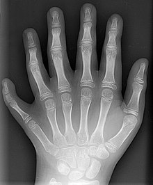
The human body can be examined with X-rays, whereby bones in particular, but also internal organs with modern devices, become visible (see also X-ray ). This takes advantage of the fact that the element calcium , which occurs in bones, with Z = 20, has a significantly higher atomic number than the elements of which the soft tissues mainly consist, namely hydrogen ( Z = 1), carbon ( Z = 6), nitrogen ( Z = 7) and oxygen ( Z = 8). In addition to conventional devices that produce a two-dimensional projection , computer tomographs are also used, which enable a spatial reconstruction of the inside of the body.
X-rays can also be used to fight cancer by targeting the cancer cells that i. A. are more sensitive to radiation than the surrounding tissue, damaged in the course of radiation therapy through targeted radiation.
Until the development of the first antimycotics , fungal skin diseases were also treated with X-rays (see also ringworm affair ).
In materials physics , chemistry , biochemistry , crystallography and other sciences, diffraction of X-rays is used for structure elucidation in the broadest sense, e.g. B. to examine the texture or for the actual crystal structure analysis . A well-known example is the elucidation of the structure of DNA . With the help of X-ray photoelectron spectroscopy (XPS), the elemental composition of a sample can be examined. In addition, XPS offers the possibility of examining chemical bonds.
In addition, the elemental composition of a substance can be determined with X-rays. The substance to be analyzed is irradiated with electrons in an electron beam microprobe (or equivalent in an electron microscope ), whereupon the atoms are ionized and emit characteristic X-rays. X-rays can also be used instead of electrons. Then one speaks of X-ray fluorescence analysis (XRF).
Discovery story
Wilhelm Conrad Röntgen is considered to be the discoverer of the rays that are named after him in German-speaking countries today, although it is certain that others before him produced X-rays. In the tubes developed by Johann Hittorf and William Crookes , which Röntgen also used for his experiments, X-rays are generated, which were detected in experiments by Crookes and from 1892 by Heinrich Hertz and his student Philipp Lenard by blackening photographic plates, but without evidently being detected to be clear about the importance of the discovery. In 1881 Johann Puluj developed a luminescent lamp, later known as the Puluj lamp, which was a prototype of an X-ray tube. Also Nikola Tesla experimented from 1887 with cathode ray tubes and thereby generated X-rays, but did not publish his results.
The first observation of X-rays by Wilhelm Conrad Röntgen took place at the Physics Institute of the Julius Maximilians University of Würzburg on the late Friday evening of November 8, 1895, when - as he himself described it - "there were no more submissive spirits in the house". Just seven weeks later, on December 28, 1895, he submitted a paper for publication under the title: About a new kind of rays . He discovered the radiation when he observed fluorescent objects near the tube during the operation of the cathode ray tube, which began to glow brightly despite the tube being covered (with black cardboard). Röntgen's achievement is to have recognized the importance of the newly discovered rays early on and to have been the first to scientifically investigate them. The x-ray of his wife's hand, which he depicted in his first publication on X-rays, certainly contributed to Röntgen's fame. After Röntgen had sent his work On a New Kind of Radiation to colleagues and friends on January 1, 1896 , including the Viennese physicist and director of the 2nd physical-chemical institute of the University of Vienna Franz Exner , from whom the Prague physicist Lechner on The news on January 4th was published on January 5th in the Vienna daily newspaper Die Presse , edited by Lechner's father. An employee of the daily brought the article to the attention of the Vienna representative of the Daily Chronicle and he immediately telegraphed it to London. From London, on the evening of January 6th, news of Röntgen's (or "Professor Routgens") discovery was telegraphed worldwide, on January 7th the London Standard printed its report on the "photographic discovery" and on January 8th this cable message became published in American magazines. Detailed reports also appeared in the Frankfurter Zeitung on January 7th and 8th. On January 9th, a note about the events was published in a Würzburg newspaper (imprecisely and without Roentgen's consent), which was the basis for further newspaper reports. In mid-January, numerous other experiments with cathode ray tubes were reported in the tabloid and specialist press. In March 1897, Röntgen published his third communication. At this time, the causal connection between cathode rays and X-rays as well as the origin of X-rays in the thin Hertz-Lenard metal foils of the Lenard tube was proven. Röntgen was honored with the first Nobel Prize in Physics in 1901 , with the Nobel Prize Committee highlighting the practical importance of the discovery.
The name X-rays goes back to the anatomist Albert von Kölliker , who on January 23, 1896 proposed the name "X-ray radiation". The occasion was Röntgen's first public lecture about his discovery, at the invitation of the Physico-Medical Society in Würzburg , which was founded by Kölliker and chaired by Karl Bernhard Lehmann . In some language areas, the name X-rays (for example English X-rays ), which was introduced by Röntgen himself, remained .
On March 26, 1896, the police, who requested permission for a lecture planned in Vienna with "Experiment with X-rays", had concerns about the use of X-rays. The nature of X-rays as electromagnetic waves was proven in 1912 by Max von Laue .
Related topics
- The N-rays supposedly discovered shortly after the X -rays turned out to be a scientific error.
- The Z machine in New Mexico is currently the most powerful X-ray source in the world.
- X-ray absorption spectroscopy
- Ankylography
literature
- Ch. R. Friedrich: 100 years of X-rays. First Nobel Prize in Physics . In: Materials Science and Technology . tape 26 , no. 11-12 , 1995, ISSN 0933-5137 , pp. 598-607 , doi : 10.1002 / mawe.19950261106 .
- Karl Heinrich Lieser: Introduction to Nuclear Chemistry . 3. Edition. Wiley-VCH, 2000, ISBN 3-527-28329-3 , pp. 143 .
- Monika Dommann : Review, Insight, Caution: A History of X-Rays, 1896–1963 . Chronos, Zurich, ISBN 3-0340-0587-3 (also dissertation at the University of Zurich , 2002).
Web links
- Basics of X-ray diagnostics ( Memento from March 6, 2008 in the Internet Archive )
- The fluorescence analysis of DNA unwinding (FADU) as a method of biological dosimetry (PDF; 2.2 MiB)
- X-ray radiation - an introduction with interactive experiments ( Memento from June 19, 2008 in the Internet Archive ) - Ulm University, archive version from June 19, 2008
- The "Glossary radiation protection" of the Jülich Research Center explains many terms from the radiation protection. It also includes many definitions and terms from laws (e.g. Atomic Energy Act ) and ordinances (e.g. X-ray ordinance ).
- X-ray public relations. DESY
Individual evidence
- ↑ Helmut Paul , Johannes Muhr: Review of experimental cross sections for K-shell ionization by light ions. Physics Reports 135 (1986), pp. 47-97. - abstract
- ↑ Köhn, C., Ebert, U. Angular distribution of Bremsstrahlung photons and of positrons for calculations of terrestrial gamma-ray flashes and positron beams. Atmosphere. Res. (2014), vol. 135-136, pp. 432-465
- ↑ Köhn, C., Ebert, U. Calculation of beams of positrons, neutrons, and protons associated with terrestrial gamma ray flashes. J. Geophys. Res. Atmos. (2015), vol. 120, pp. 1620-1635
- ↑ Kochkin, P., Köhn, C., Ebert, U., van Deursen, L. Analyzing x-ray emissions from meter-scale negative discharges in ambient air. Plasma Sour. Sci. Technol. (2016), vol. 25, 044002
- ^ Cooray, V., Arevalo, L., Rahman, M., Dwyer, J., Rassoul, H. On the possible origin of X-rays in long laboratory sparks. J. Atmos. Sol. Terr. Phys. (2009), vol. 71, pp. 1890-1898
- ↑ Köhn, C., Chanrion, O., Neubert, T. Electron acceleration during streamer collisions in air. Geophys. Res. Lett. (2017), vol. 44, pp. 2604-2613
- ↑ Marode, E., Bastien, F., Bakker, M. A model of the streamer included spark formation based on neutral dynamics. J. Appl. Phys. (1979), vol. 50, pp. 140-146
- ↑ Köhn, C., Chanrion, O., Babich, LP, Neubert, T. Streamer properties and associated x-rays in perturbed air. Plasma Sour. Sci. Technol. (2018), vol. 27.015017
- ↑ C. Köhn, O. Chanrion, T. Neubert: High-Energy Emissions Induced by Air Density Fluctuations of Discharges. In: Geophysical Research Letters. 45, 2018, p. 5194, doi: 10.1029 / 2018GL077788 .
- ↑ Manfred von Ardenne: electron microscopy physics · technology · results . Springer-Verlag, 2013, ISBN 978-3-642-47348-7 , pp. 127 ( limited preview in Google Book search).
- ↑ Wolfgang Demtröder: Experimentalphysik 3 . 3. Edition. Springer, Berlin / Heidelberg 2005, ISBN 3-540-21473-9 , pp. 243 (see in particular Fig. 7.33 in the Google Books version).
- ↑ Scientific statement on diseases caused by ionizing radiation (PDF)
- ↑ H. Schober: The direct perception of X-rays by the human visual sense . In: Vision Research . tape 4 , no. 3-4 , 1964, pp. 251-269 , doi : 10.1016 / 0042-6989 (64) 90007-0 .
- ↑ Julius Edgar Lilienfeld : The visible radiation of the Brennecks from X-ray tubes . In: Physikalische Zeitschrift . 20, No. 12, 1919, p. 280 ff.
- ↑ H. Boersch, C. Radeloff, G. Sauerbrey: About the visible and ultraviolet radiation released on metals by electrons . In: Journal of Physics . tape 165 , no. 4 , 1961, pp. 464-484 , doi : 10.1007 / BF01381902 .
- ↑ H. Boersch, C. Radeloff, G. Sauerbrey: Experimental Detection of Transition Radiation . In: Physical Review Letters . tape 7 , no. 2 , June 15, 1961, p. 52-54 , doi : 10.1103 / PhysRevLett.7.52 .
- ^ WC Roentgen: About a new kind of rays . (Preliminary communication.) In: From the meeting reports of the Würzburger Physik.-medic. Society , Würzburg 1895 ( Wikisource )
- ↑ Würzburg Roentgen Memorial Site
- ^ Heinz Otremba: Wilhelm Conrad Röntgen. A life in the service of science. A documentation with a scientific appreciation by Walther Gerlach . Franconian company printing house, Würzburg 1970, pp. 12-16.
- ↑ Erich Pirker (†): Gustav Kaiser (1871–1954) and Eduard Haschek (1875–1947): Two pioneers of medical radiology. A contribution to the verification of a controversial date. In: Würzburg medical history reports. Volume 13, 1995, pp. 97-107, here: pp. 97 and 103-105.
- ^ Heinz Otremba: Wilhelm Conrad Röntgen. A life in the service of science. 1970, p. 56.
- ↑ Werner E. Gerabek : Wilhelm Conrad Röntgen and his discovery of X-rays. In: Würzburg medical history reports. Volume 13, 1995, pp. 87-96; here: p. 91.
- ^ Heinz Otremba: Wilhelm Conrad Röntgen. A life in the service of science. 1970, p. 30.
- ^ Max von Laue: Interference phenomena in X-rays. Theoretical part by M. Laue, experimental part by W. Friedrich and P. Knipping. Presented by Arnold Sommerfeld at the meeting of the Bavarian Academy of Science on June 8, 1912 .
- ^ Heinz Otremba, Walther Gerlach : Wilhelm Conrad Röntgen. A life in the service of science. 1970, pp. 62-71.


