radiology
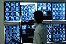
(Medical) radiology , also called radiation medicine in the narrower sense , is the branch of medicine that deals with the application of electromagnetic radiation and (with the inclusion of ultrasound diagnostics , for example) mechanical waves for diagnostic , therapeutic and scientific purposes.
In the early days of radiology, only X-rays were used and the doctrine of the application of X-rays was called radiology or radiology . In addition to X-rays, other ionizing radiation such as gamma radiation or electrons are also used. Since imaging is an essential application , other imaging methods such as sonography and magnetic resonance tomography (magnetic resonance tomography) are also counted as radiology, although these methods do not use ionizing radiation.
For diagnostic radiology include areas as part of the Neuroradiology and Pediatric Radiology. There are other areas of focus such as interventional radiology. Questions of radiation protection and the effects of radiation exposure on the human body are also of importance for the medical profession .
Radiation therapy and nuclear medicine are closely related but now independent medical specialties.
Diagnostic radiology
The imaging processes in diagnostic radiology, which has been used since 1895, include projection radiography and cross- sectional imaging processes : X-ray computed tomography , sonography and magnetic resonance tomography . In all of these processes, substances can be used that facilitate the representation or delimitation of certain structures or provide information about the function of a system. These substances are called contrast media . The selection of the procedure and the decision to use contrast media are based on the clinical question and a risk-benefit assessment. The annual costs of radiation diagnostics in Germany rose from around 4 billion euros in 1992 to more than 7 billion euros in 2008.
Radiography
In radiographic processes (also known as "conventional X-rays"), areas of the patient's body are irradiated with X-rays from one direction. On the opposite side, the radiation is registered with suitable materials and converted into an image. This shows the tissue lying in the beam path in the projection: bones absorb more radiation than soft parts and therefore cast shadows; Air-filled tissues such as the lungs are relatively permeable, so that a higher radiation intensity is registered behind them. Since various structures usually overlap in the beam path, it is often helpful to produce several images from different projection directions.
The type of sensor material used for registration depends on the device and recording type. In conventional radiography, sensitive film material analogous to photography is used, which blackens when exposed to radiation and has to be chemically developed. The semi-transparent prints can then be viewed on a light box . Further developments of this principle allow digital readout of a detector instead of the development of film material. The simplest principle is a fluorescent plate , which is scanned in after the exposure. X-ray image intensifiers are traditionally used as sensors in order to assess moving images in real time ( fluoroscopy ) . In modern devices, CCDs are used as detectors for the direct digital acquisition of both still images and real-time moving images . Radiological recordings can be saved in digital form in DICOM format.
Insoluble barium salts as suspension, iodine compounds, air and carbon dioxide are suitable as contrast media in projection radiography. Barium is commonly used for the digestive tract. Soluble iodine compounds and carbon dioxide are suitable for injection into vessels, air can be applied rectally to visualize the large intestine.
The most important examinations are listed below:
- Native = without contrast agent
- Chest X-ray: overview of the heart, lungs and chest
- X-ray skeleton
- Mammography : X-ray examination of the breast
- With contrast medium
- Angiography (representation of the vessels in general)
- Arteriography (arteries)
- Venography / venography (veins)
- Lymphography (lymph vessels)
- intravenous urography (urinary drainage system, incorrect: iv pyelogram)
- retrograde pyelography (iodine contrast medium applied to the renal pelvis via the ureter)
-
Fluoroscopy
- Contrast medium swallowing examination to visualize the esophagus
- Contrast medium meal for tracking the gastrointestinal passage
- Small intestine contrast medium examination with barium and water (double contrast )
- Colon contrast enema with barium, usually also given air (double contrast )
- Contrast examinations of the esophagus, stomach, intestines, biliary tract
- Barium contrast media ( barium sulfate , BaSO 4 ) are only administered in the digestive tract and then only if it is ensured that the contrast medium cannot escape from the digestive tract. Because when barium contrast medium enters the free body space, it encapsulates and can lead to inflammation. If barium contrast medium is inhaled into the lungs, it can lead to pneumonia .
X-ray computed tomography
See main article Computed Tomography
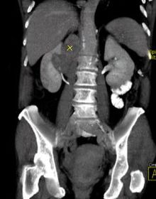
Advantages of CT: Overlay-free cross- sectional images with very high detail resolution, v. a. in bony structures, e.g. B. Inner ear. Modern devices, so-called multi-line scanners, allow the display of medium-sized and smaller vessels, e.g. B. Coronary arteries. Short recording times, with and without iodine-based contrast agent administration, also open up the gastrointestinal tract of the visual representation, so-called virtual endoscopy. Biggest disadvantage of CT: Relatively high exposure to potentially harmful X-rays, especially in the more complex examinations. This negative property of CT is particularly important in comparison to radiation-free MRI.
Magnetic resonance imaging
See magnetic resonance tomography , advantages: like CT, with better soft tissue contrast, no ionizing radiation, but higher expenditure of time and equipment, higher costs, lower tolerance in the patient v. a. Claustrophobia in conventional devices, newer design enables more open devices with good patient acceptance, contrast media for example gadolinium compounds and superparamagnetic iron oxide particles.
Ultrasound examination
See sonography , the most frequently used imaging method in medicine, advantages: gentle, repeatable, real-time assessment, sometimes functional assessment; Disadvantage: not all tissues and areas are accessible, unsuitable for very obese patients. The examination is paid too badly, so that fewer and fewer doctors are familiar with it and more and more CT and MRI examinations are used. Smallest gas bubbles (microbubbles) are used as contrast media , which facilitate the structure and function representation of vessels and the liver, as well as water and gas-absorbing substances to improve the representation of the upper abdominal organs.
education
Radiology specialist
In order to acquire the title of specialist in radiology after completing a medical degree in Germany , a five-year training period is required. The following are eligible for further training:
- 12 months in a focus area ( pediatric radiology , neuroradiology )
- 12 months in an area of immediate patient care
The content of further training to become a specialist is defined by the relevant medical associations: Proof of a certain number of independently carried out examinations in children, adults and in neuroradiology is required for admission to the specialist examination.
Statistics on this
- On January 1, 2001, 3718 Diagnostic Radiologists were registered, of which 1234 were resident. 355 did not practice any medical activity. Under the old (and now valid again) name "Radiologist", 3638 were registered, of which 1231 were resident. 1107 did not practice any medical activity.
- Together with nuclear medicine, the practice surplus averaged € 109,000 in 1998, and € 143,700 in the new federal states.
- Non-radiologists are also allowed to X-ray in Germany. In outpatient care, only about every fourth X-ray examination of those with statutory health insurance is carried out by a full-field radiologist. On the other hand, three quarters of the examinations are carried out by so-called sub-field radiologists: 32 percent of the examinations are carried out by orthopedists , in 13 percent of all cases surgeons X-ray , seven percent of the examinations are carried out by internists . The remaining examinations are carried out by doctors from other specialist groups. This is what the Federal Office for Radiation Protection (BfS) reports in its new annual report. According to this, between 2002 and 2004 every resident in Germany was x-rayed an average of 1.7 times per year. In terms of the effective radiation exposure resulting from this , the Germans, with an effective dose of 1.8 millisieverts, are "in the upper range in an international comparison," according to the BfS report. However, 50 percent of the collective effective dose can be traced back to X-ray examinations by full-area radiologists (orthopedists: twelve; internists: ten; surgeons: two percent).
Radiology technologist
In Austria, a radiology technologist is a specialist in the application of imaging processes in medicine (x-ray, sectional imaging, nuclear medicine) and for the implementation of therapeutic treatments with ionizing radiation (radiation therapy). He carries out examinations and therapies on his own responsibility as directed by a doctor, is not subject to technical instructions, is authorized to use contrast media (in cooperation with doctors) and can set up a freelance work.
In the course of the Bologna process , there was a switch to university training with an academic degree. In the winter semester of 2006, the first years started at the University of Applied Sciences Wiener Neustadt at the FH Joanneum and the University of Applied Sciences Salzburg , which finished with the bachelor's degree in summer 2008 and 2009 respectively.
In Germany, a corresponding course of study will be offered from September 2014 at the Haus der Technik in Essen .
Medical-technical radiology assistant
In Germany, radiology technologists are equivalent to medical-technical radiology assistants (MTRA). You will carry out examinations using conventional or digital radiology (e.g. CT, MRT) independently and assist with examinations such as fluoroscopy and digital subtraction angiography.
Radiographers in nuclear medicine work in the radionuclide laboratory and carry out examinations such as scintigrams, SPECT and PET. Radiotherapy technicians also participate in radiation therapy, help with radiation planning and carry out the individual therapy fractions independently. In radiation therapy, they are therapists and the intermediary between patient and doctor. Therefore, the special medical and caring moment plays a major role in this area. In X-ray diagnostics and nuclear medicine, the patient often appears only once without being noticed during the operational process. Activities in nuclear medicine and radiology tend to be more technical. The radiotherapy technicians in radiation therapy, on the other hand, accompany the tumor patient for several weeks, sometimes even for months. Therefore, they have to deal with the patient more comprehensively: with his illness, his general condition, but also with his character and his physical and mental situation. The training takes place in Germany at vocational schools or training centers. It requires a secondary school diploma and lasts three years.
At the moment, a change in training at university level is also being discussed in Germany, or a start is being made on offering the option of further academic training to MTRAs who have already completed their training with the part-time course in Medical Radiological Technology.
In Switzerland, the training is offered at higher technical schools and also lasts three years.
Interventional Radiology
The Interventional Radiology comprises minimally invasive therapeutic measures to be carried out under permanent control means of imaging methods: for example, the distention of vasoconstriction ( angioplasty ) under fluoroscopic control (angiography). When using a vascular prosthesis (stent), this method is known as stent angioplasty . Further interventional radiology measures include: a .: tumor embolizations (~ sclerosing), the treatment of acute bleeding, elimination of duct stenosis in the gastrointestinal tract or in the biliary tract, tissue removal and the treatment of vascular dilatations (aneurysms) Interventional radiology is systematically not part of diagnostic radiology, but historically evolved from it and is mostly carried out by radiologists.
Radiation protection
Since the radiation doses used in X-ray diagnostics are very low, but potentially harmful for the patient and the user, special emphasis is placed on radiation protection in radiology . The German Society for Medical Radiation Protection is an association of doctors and other competent persons who have set themselves the goal of exploring and minimizing these radiation risks in medicine.
With around 1.3 x-rays per inhabitant per year, Germany takes a top position. The medical application of ionizing radiation leads to an additional radiation exposure of roughly 2 mSv / a per inhabitant. Theoretically 1.5% of the annual cancer cases can be traced back to this.
Computed tomography has by far the largest share of medical radiation exposure .
A basic guideline for minimizing radiation exposure when using radiological methods is published by the working group "Orientierungshilfe Radiologie", the Federal Radiology Group of the Austrian Medical Association and the Austrian Radiological Society , as a non-binding reference work both in paper form and online. The German Radiation Protection Commission also offers such guidance.
literature
- W. Angerstein (Ed.): Fundamentals of radiation physics and radiological technology in medicine. 5th edition. H. Hoffmann Verlag, 2005.
- Roland C. Bittner : Radiology Guide. ISBN 3-437-41210-8 , KNO 06 29 50 87.
- Martin Breitenseher, Peter Pokieser, Gerhard Lechner: Textbook of radiological-clinical diagnostics. 2nd Edition. University Publisher 3.0, 2012. ISBN 978-3-9503296-0-5 .
- Susanne Hahn: Radiology. In: Werner E. Gerabek , Bernhard D. Haage, Gundolf Keil , Wolfgang Wegner (eds.): Enzyklopädie Medizingeschichte. De Gruyter, Berlin / New York 2005, ISBN 3-11-015714-4 , p. 1259 f.
- Susanne Hahne: Radiology. In: Encyclopedia of Medical History. 2005, p. 1212.
- Dirk Pickuth: Radiology facts. Uni-Med, Bremen 2002, ISBN 3-89599-310-7 , KNO-NR: 11 11 20 48.
- Jörg-Wilhelm Oestmann: Radiology. A case-based textbook. Thieme, Stuttgart 2002, ISBN 3-13-126751-8 , KNO-NR: 10 91 20 07.
- Theodor Laubenberger, Jörg Laubenberger: Technology of medical radiology. Diagnostics, radiation therapy, radiation protection. For doctors, medical students and MTRA. Deutscher Ärzte-Verlag, ISBN 3-7691-1132-X , KNO-NR: 00 99 81 31.
- German Röntgen Museum (ed.): The eyes of the professor. Wilhelm Conrad Röntgen. A short biography. Past Publishing, Berlin 2008.
- Klaus Wicke, Franz Frühwald, Dimiter Tscholakoff ( Austrian X-ray Society ) and Franz Kainberger: Orientierungshilfe Radiologie - Instructions for the optimal use of clinical radiology 4th edition. 2011, ISBN 978-3-902552-99-0 , online version: http://orientierungshilfe.vbdo.at/
Magazines
- RöFo, Advances in the field of X-rays and imaging processes , organ of the German and Austrian X - Ray Society , Thieme-Verlag , ISSN 0936-6652
- The Radiologist , Springer Verlag, ISSN 0033-832X
- radiology assistant , Schmidt-Römhild Verlag, ISSN 0935-1779
See also
- Computer-assisted detection
- Digital X-ray
- X-ray screening
- X-ray sign
- Medical technology
- History of radiation protection
Web links
- Cardiovascular and Interventional Radiological Society of Europe
- German Radiological Society
- European Society of Radiology
- radiopaedia.org (Radiology Wiki)
- hellste-koepfe.de - Portal for and about radiologists
- Orientation aid radiology - Instructions for the optimal use of clinical radiology
- Medicine with a perspective - information initiative by radiologists and radiation specialists
Individual evidence
- ↑ Federal Statistical Office 2010, quoted from Apotheken-Umschau, July 1, 2010, p. 57
- ↑ Quoted from: Ärzte Zeitung, August 21, 2008, partial radiologists x-ray in three out of four cases .
- ↑ Studies. (No longer available online.) Association of Radiotechnologists Austria, archived from the original on December 17, 2015 ; Retrieved November 26, 2015 . Info: The archive link was inserted automatically and has not yet been checked. Please check the original and archive link according to the instructions and then remove this notice.
- ↑ Bachelor's degree in Medical Radiological Technology, part-time , Haus der Technik, accessed on August 5, 2014
- ^ Rolf Sauer: Radiation Therapy and Oncology. 5th edition, Urban & Fischer, p. 15.
- ↑ Bachelor's degree in “Medical Radiology Technology” starts in Essen in September ( memento of the original from April 15, 2015 in the Internet Archive ) Info: The archive link has been inserted automatically and has not yet been checked. Please check the original and archive link according to the instructions and then remove this notice. , Interview on MTA-Dialog.de, accessed on August 5, 2014
- ↑ Medical-technical radiology - MTR specialist specialist in medical-technical radiology. Retrieved January 11, 2019 .
- ↑ de Gonzalez and Berry, Lancet 2004; 363: 345-51.
- ^ De Gonzalez, Sarah Darby: Risk of cancer from diagnostic X-rays: estimates for the UK and 14 other countries. Lancet 2004; 363: 345-51, doi: 10.1016 / S0140-6736 (04) 15433-0 .
- ↑ Orientation aid for imaging examinations , recommendation of the Radiation Protection Commission (PDF; 566 kB)


