Ankle joint

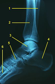
Legend:
1 - fibula (fibula)
2 - shinbone (tibia)
3 - ankle bone (talus)
4 - heel bone (calcaneus)
5 - scaphoid (navicular bone)
6 - metatarsal bone (Os metatarsi)
The ankle is the joint between the lower leg and the foot . A distinction is made between the upper ankle joint (OSG) and the lower ankle joint (USG). Both ankle joints together are functionally a cylinder joint ( Articulatio cylindrica ).
Upper ankle joint (ankle)
Ankle ( lat. Articulatio talocruralis ; also ankle joints ) are the lower ( distal ) ends of the tibia ( tibia ) and the fibula ( fibula ) and the ankle bone ( talus ) the articulating bones. In detail, these are defined by the inner ankle ( medial malleolus ) of the tibia and outer ankle ( lateral malleolus ) of the fibula formed ankle mortise (also ankle joint ) and the talus pulley ( trochlea tali ).
The malleolar fork is held together by the distal tibiofibular syndesmosis . Syndesmotic injuries are found in around 16% of all ankle injuries and are typically also present in Weber C and sometimes Weber B fractures of the outer ankle . A distinction is made between four bands :
- The interosseous tibiofibular ligament is the distal part of the interosseous membrane and can be separated from it as an independent ligament. The fibers run obliquely downwards from the tibia to the fibula in a lateral-distal-anterior direction. The ligament is about 2-3 cm wide and ends about 1 cm above the joint space. The ligament contributes about 22% to the stability of the syndesmotic apparatus.
- The anterior tibiofibular ligament (anterior syndesmotic ligament) runs in an oblique direction from the clearly delimited anterior tubercle ( Tubercule de Chaput Tillaux ) on the lateral distal edge of the tibia to the anterior tubercle of the fibula. It contributes about 35% to the syndesmosis stability.
- The posterior tibiofibular ligament (posterior syndesmotic ligament) connects the posterior edge of the lateral malleolus with the posterior tubercle on the lateral posterior edge of the tibia and is much more horizontal than the anterior syndesmotic ligament. It has a share of about 9% in the syndesmosis stability.
- The transverse tibiofibular ligament was described partly as a separate ligament, partly as the deeper and distal part of the posterior syndesmotic ligament and also has a horizontal orientation. After the anterior syndesmosis, it contributes significantly to the stabilization of the syndesmosis with about 33%.
The mechanics of the upper ankle joint does not quite correspond to a hinge joint , because its axis runs diagonally through the ankle roll and the ankle joint fork. This allows - described simplified - the reduction ( plantar flexion ) and the lifting of the foot (dorsal flexion or dorsiflexion ). In addition, a small amount of internal and external rotation as well as an inward ( pronation ) and minimal outward rotation ( supination ) of the ankle roll is possible. According to the neutral-zero method , the range of motion (plantar-dorsal) thus covers a range of 50 ° –0 ° –30 °. However, the ankle roll does not have the full shape of a cylinder , as it is wider at the front than at the back. The slight movements to the side ( abduction and adduction ), which are greater when the foot is lowered, are hardly possible when the foot is raised. The ankle joint is one of the most heavily stressed joints in the body, as it has to carry the entire body load with every step and transfer it to the ground.

This high load and the incompletely cylindrical anatomy as well as the different design of ligament connections result in a wide range of injuries, which primarily affect the ligaments, but also the bones. Ankle injuries are extremely common. " Sprains " and " twisting " of the joint are particularly common.
In the ankle joint, without previous injury, signs of wear ( arthrosis ) develop less often than in other joints . Most of the arthroses of the ankle are secondary (post-traumatic) arthrosis, i.e. the long-term consequences of severe or inadequately treated injuries such as ankle sprained fractures or complex capsule ligament injuries . Developments and techniques that are fully developed on the knee joint to improve the cartilage surface (transplantation of cartilage cells or cartilage-bone cylinders) or to replace it with an endoprosthesis (artificial joint) are now also successful in the ankle. The treatment options are still limited, especially because the joint is irregular and very narrow and therefore not as easily accessible to a variety of arthroscopic treatments as the knee joint, for example. Basically, in many cases, a graduated therapy scheme is used and conservative treatment (including technical shoe measures) is started, which in some cases leads to years of relief from the symptoms. If these possibilities are exhausted when osteoarthritis develops, there are essentially two surgical therapy options: the stiffening of the upper ankle joint ( arthrodesis of the ankle joint ) or the installation of an artificial joint ( ankle joint endoprosthesis ).
Lower ankle joint (USG)
The lower ankle ( lat. Articulatio talotarsalis ) is part of the foot and is itself in turn in the front lower ( Articulatio talocalcaneonavicularis ) and the lower rear ankle joint ( articulatio subtalar joint , articulatio talocalcanearis divided). The two chambers are separated by the ankle-calcaneus ligament ( ligamentum talocalcaneum interosseum ), this ligament runs in the canalis tarsi . The main function of this ligament is to guide blood vessels to nourish the talus .
The posterior chamber of the lower ankle joint ( articulatio subtalaris ) is formed by the ankle bone (talus) and heel bone (calcaneus). The posterior articular surface of the calcaneus ( Facies articularis calcanea posterior ) and the posterior articular surface of the ankle bone ( Facies articularis talaris posterior ) are connected here. The anterior chamber of the lower ankle joint ( articulatio talocalcaneonavicularis ) is formed by the ankle bone, calcaneus and scaphoid bone ( navicular bone ). Here, the anterior and middle joint surfaces of the heel and talus articulate with one another. Another articulated connection, the talonavicular joint, exists between the ankle bone and the navicular bone; the corresponding joint surfaces of the talar bone ( facies articularis talaris ) and the navicular bone ( facies articularis navicularis ) are articulated. The heel and navicular bone together form a hollow in which the talus has only one axis of movement.
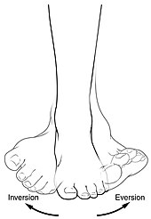
This axis of movement of the lower ankle joint runs from the center of the scaphoid to the outside of the calcaneus. The angle to the horizontal plane is approximately 30 °, the angle to the sagittal plane is approximately 20 °. In the lower ankle, only two movements are possible: 20 ° eversion (lifting the outside of the foot) and 35 ° inversion (lifting the inside of the foot) from the neutral position .
Ligaments of the ankle
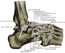
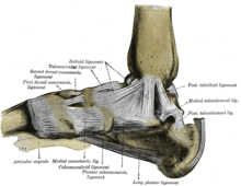
The ankle is held together by a series of ligaments. The deltoid ligament ( Ligamentum deltoideum or Ligamentum collaterale mediale ) consists of a shin-scaphoid bone part ( pars tibionavicularis ), a shin-bone- calcaneus part ( pars tibiocalcanea ) and an anterior and posterior shinbone-ankle bone part ( pars tibiotalaris anterior and posterior ) . The lateral ligament ( Ligamentum collaterale laterale ) is formed by the anterior and posterior talofibular ligament ( Ligamentum talofibulare anterius and Ligamentum talofibulare posterius ) as well as a heel bone-fibula ligament ( Ligamentum calcaneofibulare ). The ankle joint is held together by the anterior and posterior tibial-fibrous ligaments ( ligamentum tibiofibular anterius and ligamentum tibiofibular posterius ). Between the neck of the talus and the dorsal surface of the scaphoid lies the talonavicular ligament .
The outer ligaments are particularly often affected by twist injuries; in this case one speaks of an outer ligament rupture . Ankle injuries often lead to damage to the capsular ligament apparatus (ligament strain, stretching, tearing). Bony injuries are rare (fracture of the outer and inner ankles, rupture of the connecting ligament between the tibia and fibula). The ankles are very often affected, accounting for around 20% of all sports injuries.
literature
- H. Ferner, J. Staubesand: Sobotta. Atlas of Human Anatomy. 18th edition. Urban & Schwarzenberg, 1982, ISBN 3-541-02828-9 .
- Herbert Lippert : Textbook anatomy. 8th edition. Elsevier, 2011, ISBN 978-3-437-42365-9 .
Web links
Individual evidence
- ^ KE McKeon, RW Wright, JE Johnson, JJ McCormick, SE Klein: Vascular anatomy of the tibiofibular syndesmosis. In: Journal of Bone and Joint Surgery. [On] 2012; Volume 94-A, pp. 931-938.

