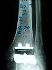
Ankle joint endoprosthesis
history
The upper ankle prosthesis is not a new invention. Artificial ankles have been used since around 1970. The optimistic expectations from the beginning could not be confirmed in the long-term observation. The prostheses loosened after a few years and had to be removed: the design and anchoring method of the first-generation prostheses were not yet fully developed. In the meantime, however, there has been experience with modern total endoprostheses of the ankle for around 20 years .
Components
The modern ankle joint endoprostheses consist of three components:
- a rounded cap for the talus roll made of a cobalt-chrome alloy,
- a plate for the tibial articular surface made of a cobalt-chromium alloy, and
- a freely movable polyethylene sliding core or attached to the tibia component.
The stability in the artificial joint is adjusted via the height of the polyethylene sliding core.
The cement-free anchoring of the two components on the ankle bone (talus) and shin bone (tibia) resulted in a decisive improvement in the modern ankle joint endoprosthesis . A special coating, for example with titanium / calcium phosphate, enables the bone to grow firmly together with the implants.
Types of prostheses and surgical technique
There are currently several prosthesis models, in 2- and 3-component designs. Most ankle prostheses are inserted through an anterior, longitudinal access to the upper ankle. A sufficiently long skin incision is made to avoid pulling the hook on the soft tissue. Using precise alignment and sawing templates, the bone bed in the area of the tibia and talus is appropriately trimmed so that the prosthesis can be inserted. Methods have existed since 2015 to have the cutting blocks for resection on the tibia and talus created individually for each patient using CT data in order to enable precise saw cuts. Here, the alignment is determined preoperatively and a computer creates a saw guide that is precisely adapted to the bone, which saves the surgeon from having to assess alignment angles and resection lines during the operation.
The procedure is usually performed under regional anesthesia , in which either the lower half of the body or only the affected leg are included in the anesthesia . The main advantage of this method is that it makes pain therapy easier after the operation. In special cases, the procedure is performed under general anesthesia and takes between 90 and 120 minutes. Immediately after the operation, the patient is provided with a removable Vacoped® or DARCO posterior splint. If the course is uncomplicated, the patient can get up on the 1st day after the operation with help. On the 2nd day after the operation, the leg is fully axially loaded in a controlled manner with the Vacoped® splint. This is used to recompress the prosthesis components in the bone bed and to immobilize the ankle joint and the scar postoperatively.
indication
In order to find out which patients are suitable for a prosthesis, a thorough physical examination of the patient, which includes an X-ray of the diseased joint, is carried out before each operation. A magnetic resonance imaging (NMR) can be a necessary additional investigation in certain cases. Ankle replacement is not possible with circulatory disorders in the ankle area, infections and severe soft tissue problems in the ankle area. Gross malpositions in the ankle joint can make the operation considerably more difficult. The implantation of an artificial ankle is a technically demanding and difficult operation. It should therefore be carried out by experienced surgeons who are familiar with this problem. In Germany, around 850 ankle prostheses are currently implanted each year. The centers with the greatest experience in the field of ankle arthroplasty are currently in Magdeburg, Basel , Endoklinik Hamburg , Berlin , Munich , Offenburg , Weilheim / Bavaria , Wiesbaden and Zurich .
Complications
As with other surgeries, complications can arise with the implantation of an ankle prosthesis. A fracture of the inner or outer ankle either during the operation or immediately after the operation must then be surgically stabilized. Wound healing disorders and other soft tissue problems sometimes require longer treatment, and occasionally additional plastic surgery. In some cases, follow-up operations must also be performed. The reason for this can be, for example, in addition to broken bones, the loosening of individual prosthesis components, persistent pain, or too much restricted mobility. A replacement of prosthesis parts or a stiffening operation is rarely necessary. Because of the relatively low bone loss with the newer prosthesis models, the stiffening operation ( arthrodesis of the ankle joint ) is still possible as a withdrawal without major difficulties.
After the operation

From the 2nd day after the operation, the walking exercises with the Vacoped ® splint begin with a 20 kg partial load on the operated leg for a total of 6 weeks after the operation. In addition, from this day onwards, what is known as “interim mobilization” of the operated ankle joint with lifting and lowering of the foot is performed. In addition, there is passive exercise of the joint with a motorized splint. Manual lymph drainage and elevation promote the swelling of the joint. After the wound has healed, the threads are usually removed on the 12th day after the operation. Further physiotherapeutic treatment can then be carried out either in an inpatient follow-up treatment or an outpatient physiotherapy . After 6 weeks postoperatively, the Vacoped® splint can be removed and you can quickly switch to full load. The rehabilitation phase lasts around 12 weeks.
Check-ups
The first x-ray check is carried out immediately after the operation. The other X-ray controls should be carried out upon discharge from inpatient treatment and 6 weeks postoperatively, 12 weeks postoperatively, 6 months postoperatively and then annually as part of the clinical control examinations.
literature
- B. Hintermann: The STAR ankle prosthesis. Short and medium term experiences. In: Orthopedist. 28, 1999, pp. 792-803.
- H. Kofoed, J. Stürup: Comparison of ankle arthroplasty and arthrodesis. A prospective series with long term follow-up. In. Foot. 4, 1994, pp. 6-0.
- H. Kofoed: The development of ankle arthroplasty. In: Orthopedist. 28, 1999, pp. 804-811.

