Ankle bone
The ankle bone (syn. Roll bone , med. Talus , obsolete also astragalus ) is a short bone and part of the tarsus and the ankle . It lies between the ankle fork ( malleolar fork ) and heel bone ( calcaneus ) and connects the foot with the leg.
construction
The talus consists of the body ( corpus tali ), the neck ( collum tali ) and the talus head ( caput tali ).
On the top of the talus is the talus roll ( trochlea tali ). In the side view, the talus roll is convex, with a greater curvature at the front than at the back. In the front view, however, there is a concavity, the roll is arched in the middle with the prominent side edges and matches the slight central protrusion of the end of the shin. This also secures and guides the shin roll in the ankle fork. The back of the shin roll is somewhat narrower and less arched, so more tilting movements are possible in the upper ankle when the foot is flexed. On the other hand, the tibial roll is slightly wider at the front than the ankle fork and therefore fits securely in the normal stance with stretching of the tibiofibular syndesmosis . This leads to a high joint stability of the upper ankle joint in normal stance.
Medial to the trochlea tali is the comma-shaped joint surface for the medial malleolus ( Facies malleolaris medialis ). The posterior tali process descends from this articular surface . Lateral to the trochlea tali is the triangular joint surface for the outer malleolus ( Facies malleolaris lateralis ). The lateral process of the talus branches off from this articular surface .
The joint surface for the scaphoid bone ( navicular bone ) is located on the head of the talus . This joint surface is known as the articularis navicularis face . Here the spherical head lies in the concave articular surface of the scaphoid bone, which forms the talonavicular joint. The articular surface is part of the anterior division of the lower ankle joint (USG).
On the back there is a pronounced extension, the processus posterior tali . This is divided by a groove (sulcus) through which the tendon of the long flexor of the big toe runs ( sulcus tendinis musculi flexoris hallucis longi ). The lateral part ( tuberculum laterale processus posterioris tali ) is much more prominent and, as the os trigonum, can represent an independent accessory bone , which is found in 13% of people. The posterior part of the external ligament inserts into the lateral part.
On the underside are the following three articular surfaces that are in contact with the calcaneus:
- Facies articularis calcanea anterior
- Facies articularis calcanea media
- Facies articularis calcanea posterior
Between the facies articularis calcanea media and the facies articularis calcanea posterior there is a furrow known as the sulcus tali . Together with a corresponding furrow in the calcaneus ( sulcus calcanei ), this furrow forms the sinus tarsi .
In Paarhufern the talus has two castors. The upper joint roller ( Trochlea tali proximalis ) forms the articulated connection to the joint screw of the tibia ( Cochlea tibiae ) and the lower joint roller ( Trochlea tali distalis ) articulates with the Os tarsi centrale and quartum of the ankle.
Faulty investments
Malformations:
- Tarsal coalitions Coalitio , synostoses (fusion) with the calcanaeus and / or the os naviculare tarsi
- Talus cleft , incomplete fusion of the embryonic nuclear structures
- Globular talus , congenital misalignment with a corresponding change in the upper ankle
Malpositions:
- Talus verticalis
- Talus obliquus
Fractures
Broken bones (fractures) of the ankle bone are very rare and account for only 0.32% of all bone fractures and only 3.4% of all fractures in the foot. Ankle fractures are virtually non-existent in children. The causes are usually falls from a great height or accidents with high energy. The first case series were described during the world wars and mainly concerned pilots who had jumped off and parachutists, from which the name aviator's astragalus ( aviator's talus ) comes from.
The most common are the talar neck fractures between the distal talus head and the proximal talar roll. This usually requires forced dorsiflexion of the foot, usually combined with a supination or pronation movement (which is why accompanying ankle fractures are not uncommon). The Hawkins classification is very often used for talar neck fractures:
- ° 1 - unshifted talar neck fracture
- ° 2 - Displacement with dislocation of the head of the talus together with the calcaneus against the roll of the talus
- ° 3 - The proximal fragment (the talus roll) dislocates completely backwards
- ° 4 - Both fragments are luxated, the proximal fragment backwards and the distal fragment at least partially anteriorly.
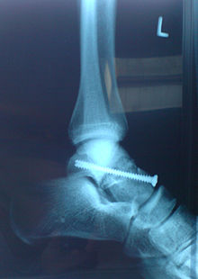
Treatment of a fracture of the ankle bone depends on the location and severity of the fracture. In the case of a fracture without moving the ends of the fracture, the foot is usually immobilized in a lower leg cast . If the ends of the fracture are displaced (dislocated), an operative method ( osteosynthesis ) is usually used. The risk of a circulatory disturbance with subsequent osteonecrosis , especially of the talus body, is very high with displaced or multi-fragment fractures, so a long relief phase is necessary after the operation. A fracture of the lateral process of the talus, known as a snowboarder's ankle , can occur, especially in snowboarders .
An os trigonum on the back of the ankle bone is an accessory bone that arises due to its constitution or can be the result of an avulsion fracture of the tuberculum laterale processus posterioris tali , by pulling the posterior part of the lateral ligament ( ligamentum talofibulare posterius ) inserted there in supination injuries of the upper ankle joint .
In the past, a more frequently used procedure for multi-fragment fractures of the ankle bone was its complete removal ( astragalectomy ). This leaves an often painful restriction of movement with a disruption of normal foot biomechanics. In general, the most anatomical reconstruction possible is sought today.
literature
- ^ A b c d e Titus von Lanz , Werner Wachsmuth : Practical anatomy - leg and statics (first volume, fourth part). Springer-Verlag Berlin, 2004 (2nd edition, special edition, ISBN 3-540-40570-4 )
- ^ Franz-Viktor Salomon: Bony skeleton. In: Salomon, F.-V. u. a. (Ed.): Anatomy for veterinary medicine. Enke-Verlag, Stuttgart 2004, pp. 37-110. ISBN 3-8304-1007-7
- ↑ A. Rozansky, E. Varley, M. Moor, DR Wenger, SJ Mubarak: A radiologic classification of talocalcaneal coalitions based on 3D reconstruction. In: Journal of children's orthopedics. Volume 4, number 2, April 2010, pp. 129-135, doi: 10.1007 / s11832-009-0224-3 , PMID 20234768 , PMC 2832879 (free full text).
- ↑ Kind-und-Radiologie ( Memento of the original from March 4, 2016 in the Internet Archive ) Info: The archive link was inserted automatically and has not yet been checked. Please check the original and archive link according to the instructions and then remove this notice. (PDF)
- ^ W. Blauth, W. Harten, A. Kirgis: Frontal Talusspalte - Talus bipartitus . In: Zeitschrift für Orthopädie und their Grenzgebiete , 125, (1987), pp. 302-307, ISSN 0044-3220 ; CODEN ZOIGAP
- ↑ F. Hefti: Pediatric Orthopedics in Practice . Springer 1998, ISBN 3-540-61480-X
- ↑ Jens Vanbiervliet, Kris Van Crombrugge, Jan ML Bosmans, Filip Vanhoenacker: Un sévère traumatisme du tarse . In: Ortho-Rhumato , 2013, Volume 11, Issue 6, December 2013, pp. 32–35


