Os trigonum

The os trigonum is an additional (accessory) bone of the talus . After the Os tibiale externum, it is the most common and most important accessory bone of the foot skeleton. Injuries and repeated microtraumas - especially in athletes - can lead to problems with the os trigonum (hindfoot problems), which are known as the os trigonum syndrome .
description
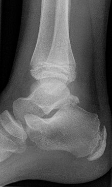
The os trigonum arises from a separate bone core that is located on the posterior edge of the talus ( talus ). In girls, this usually happens between the ages of 8 and 10 and in boys between 11 and 13. Within a year, the core of the bone usually joins the main bone.
Incidence
About 3 to 15 percent of all adults have an os trigonum, and about 1.4 percent have it on both feet. In a radiological study carried out in Japan with 3460 x-rayed feet, an os trigonum was found in 438 feet (= 12.7%) .
Os trigonum syndrome
From an anatomical point of view, the os trigonum is located in the vicinity of the flexor hallucis longus muscle (long flexor of the big toe), the posterior fibulotalare ligament (a ligament that leads from the fibula head backwards and downwards to the ankle) and the deltoid ligament (the inner ligament of the ankle, deltoid ligament). Strong tensile stress or stretching of the two ligament structures, as well as excessive tensile stress on the tendon of the long big toe flexor, can lead to irritation of the os trigonum . In such cases, the affected patients usually complain of stress-dependent, permanent pain that they feel behind the outer ankle. Add to this the feeling of stiffness and weakness in this area. The pain can also be caused by a tear in the posterior process or a pseudarthrosis .
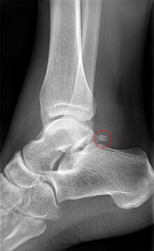
The X-ray shows the trigonum as a triangular, round or oval structure on the posterior tali process when viewed from the side. In most cases it is a single bone, but several os trigonum can also be formed.
In most cases the os trigonum syndrome is initially treated conservatively. This includes the local administration of cortisone (as an injection) or the infiltration of a local anesthetic . Oral use of nonsteroidal anti-inflammatory drugs can also help relieve pain. On the physical therapy side , stretching exercises and massages can be helpful. More cushioned shoes (air cushions) are often recommended to those affected. For competitive athletes, modified training can also bring relief. If the conservative methods do not bring significant relief for the patient, the surgical removal of the os trigonum remains the last resort . In top athletes, the foot is fully resilient two to three months after the operation.
Initial description
The os trigonum was first described in 1804 by the German surgeon Johann Christian Rosenmüller on the occasion of his inaugural lecture as Professor of Anatomy and Surgery in Leipzig.
Individual evidence
- ↑ J. Steinhäuser: Differentiation disorders and variations of the foot skeleton. In: Orthopedics in practice and clinic. A. Witt et al. (Editor) Verlag Thieme, 1985, ISBN 3-135-61902-8 , pp. 2.1-2.27. limited preview in Google Book search
- ↑ a b c d J. Characters including: The Os trigonum syndrome. In: Der Unfallchirurg 102, 1999, pp. 320–323. doi : 10.1007 / s001130050409
- ^ CJ Wirth: Orthopedics and orthopedic surgery. Foot. Georg Thieme Verlag, 2002, ISBN 3-131-26241-9 , p. 238f limited preview in the Google book search
- ^ RP Johnson et al.: The os trigonum syndrome: use of bone scan in the diagnosis. In: The Journal of Trauma 24, 1984, pp. 761-764. PMID 6236302
- ^ J. Lawson: International Skeletal Society Lecture in honor of Howard D. Dorfman. Clinically significant radiologic anatomic variants of the skeleton. In: AJR 163, 1994, pp. 249-255. PMID 8037009 (Review)
- ↑ T. Tsuruta et al.: [Radiological study of the accessory skeletal elements in the foot and ankle.] In: Nippon Seikeigeka Gakkai Zasshi 55, 1981, pp. 357-370. PMID 7276670 (article in Japanese, abstract in English)
- ^ AE Brodsky and MA Khalil: Talar compression syndrome. In: Am J Sports Med 14, 1986, pp. 472 ± 476. PMID 3799872
- ^ AE Brodsky and MA Khalil: Talar compression syndrome. In: Foot Ankle 7, 1987, pp. 338-344. PMID 3609985
- ↑ N. Maffulli et al .: Traumatic lesions of some accessory bones of the foot in sports activity. In: J Am Podiatr Med Assoc 80, 1990, pp. 86-90. PMID 1968098
- ↑ a b J. Wenig: Os trigonum syndrome. In: J Am Pod Med Ass 80, 1990, pp. 278-282. PMID 2366175
- ^ W. Hamilton: Surgical anatomy of the foot and ankle. In: Clin Symposia 37, 1985, pp. 1-32. PMID 4064598
- ↑ B. Veazey et al.: Excision of ununited fractures of the posterior process of the talus: a treatment for chronic posterior ankle pain. In: Foot Ankle 13, 1992, pp. 453-457. PMID 1483605
- ^ A. Prescher: Proceedings on the 10th Teratology Symposium Greifswald, September 23-25, 1999. R. Vogel (editor), Tectum Verlag, 2001, ISBN 3-828-88248-X limited preview in the Google book search
literature
- RL Blake et al: The os trigonum syndrome. A literature review. In: J Am Podiatr Med Assoc 82, 1992, pp. 154-161. PMID 1578352 (Review)
- DM Jones, CL Saltzman, G. El-Khoury: The diagnosis of the os trigonum syndrome with a fluoroscopically controlled injection of local anesthetic. In: The Iowa orthopedic journal. Volume 19, 1999, pp. 122-126, PMID 10847526 , PMC 1888622 (free full text).
- D. Karasick and ME Schweitzer: The os trigonum syndrome: imaging features. In: AJR Am J Roentgenol 166, 1996, pp. 125-129. PMID 8571860

