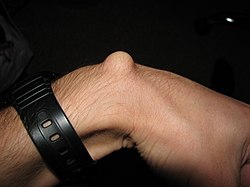
Ganglion (upper leg)
| Classification according to ICD-10 | |
|---|---|
| M67.4 | Ganglion of a joint or tendon (s) - (sheath) |
| ICD-10 online (WHO version 2019) | |


A ganglion (colloquially, like other similar-looking phenomena, imprecisely also referred to as the fuselage ) is a single or multiple, benign tumor formation in the area of a joint capsule or superficial tendon sheath (tendon sheath hygroma). The term “overbone” is misleading insofar as the use of the term “leg” suggests that a bone is affected (see nasal bone , heel bone , shin bone ). A real over leg is called an exostosis .
causes
The reason for the formation is mostly unknown; Overuse of the corresponding structures with chronic irritation, but also spontaneous formation, are assumed. Typically, a ganglion starting from a joint capsule always has a small, pedicled connection to the interior of the joint, through which an exchange of fluid between the two structures is basically possible. If, for example, there is a joint effusion as part of an activated osteoarthritis , the state of tension in the joint capsule changes - depending on the position of the joint - i.e. the internal pressure in the joint, and a ganglion can fill up more or less. In the area of joints, a ganglion can also emerge as a protuberance of the inner leaf of the joint capsule (synovial membrane) through weak points in the outer (fibrous membrane).
Occur
A ganglion is a common, benign tumor on the extensor or flexor side of the wrist or finger joints (ring ligament ganglia). The ganglion is more rarely located on the foot (back of the foot) or the knee, in individual cases also in the area of the elbow or shoulder.
Symptoms
As a resilient expulsion of a joint capsule or a tendon sheath filled with viscous fluid ( mycin ) , a ganglion can cause pain or limit mobility. In the case of very large ganglia, compression of the nerves and vessels can occur. If it occurs at the outermost, last finger joint , the nail can be deformed due to the mechanical pressure of the ganglion on the nail root . This type of ganglion is also called a dorsal cyst.
diagnosis
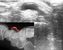
The diagnosis of a ganglion can usually be made by its location or shape. The overlying skin can be moved, there is an unchangeable connection to the joint or the tendon sheath. However, since other changes can also present a similar picture, it is usually necessary to secure the diagnosis. This can be done, for example, by needle aspiration of the liquid, ultrasound or surgical intervention. A ganglion is not visible on a normal x-ray . However, the X-ray serves to rule out a protruding bone ( exostosis ). Such an exostosis can occur on the back of the hand, especially at the base of the third metacarpal bone , which, as a carpal boss , can be confused with a ganglion.
therapy
Basically, any therapy should be discussed with the doctor, otherwise joint damage can occur. We strongly advise against the historical "treatment method" of smashing the ganglion with the blow of a Bible or a wooden hammer!
Initially, in the cases that cause less discomfort, an attempt can be made to immobilize the affected region, which often causes the ganglion to recede, but if it is again overstrained it usually occurs again (e.g. because joint effusion increases again).
Manual compression of the ganglion can also be successful. The ganglion can be massaged with moderate pressure (fluid is pushed back into the joint) or burst with strong point pressure. In some cases, this form of therapy can lead to complete healing.
Another form of therapy is the puncture of the ganglion with aspiration (suction) of the contents. Often, however, the interior of the ganglion fills up again after a while, so that surgical rehabilitation is usually the goal.
The operation can be carried out conventionally or endoscopically. In conventional surgery, the ganglion and its pedicle are removed through the smallest possible access / incision (depending on size). If the operation is carried out endoscopically, the instruments are introduced through two or three small incisions in the skin. With both surgical procedures, the disease can recur ( relapse ) in 20 to 30 percent of cases .
See also
Web links
Individual evidence
- ↑ N. Scholz: Textbook and picture atlas for podiatry. Verlag Neuer Merkur, 2007, ISBN 978-3-937346-40-3 , p. 159. (online)
- ^ W. Platzer: TaschenAtlas Anatomie. Georg Thieme Verlag, 2005, ISBN 3-13-492009-3 , p. 24. (online)
- ↑ A. Bardeleben : Textbook of surgery and operation theory. Verlag G. Reimer, 1859, p. 831. pdf
- ↑ https://www.netdoktor.de/krankheiten/ganglion/
