Bone healing
The bone healing , including fracture healing called, after a bone fracture , depending on the type of fracture ( fracture done in two ways), and the medical care: as primary and as secondary bone healing.
history
Albrecht von Haller recognized the importance of blood vessels for fracture healing as early as 1767 based on an experimental investigation.
Primary bone healing
Primary bone healing can only take place if the fracture ends are closely adapted as early as possible after the fracture and they cannot move relative to one another. This is usually only the case with surgical treatment using osteosynthesis .
No external callus is formed during primary bone healing . The trabeculae ( substantia spongiosa ) grow together through the accumulation of newly formed bone tissue. This bone tissue is formed by activating the osteoblasts of the inner periosteum ( endost ). In the area of the marrow cavity of the long bones , an internal bone callus usually forms from trabeculae. The osteons of the compact bone substance ( substantia compacta ) at the two ends of the fracture can, with very good adaptation (gap <300 µm), grow together from both ends of the fracture and fuse again. The newly emerging bone tissue initially has a lower mechanical load capacity. It is then broken down again by osteoclasts from about the 8th week and replaced by bone tissue aligned with the pressure and tensile load lines of the bone (trajectories) (so-called remodeling ).
Secondary bone healing
A secondary bone healing occurs with less good adaptation and fixation of the broken ends, as well as in larger bone defects - such as after a comminuted fracture - on.
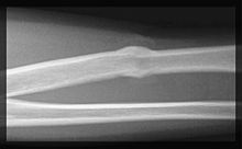
First, blood comes out of the fracture surface and a bruise ( hematoma ) forms. This leads to an activation of the inflammatory cascade and inflammatory cells release cytokines such as interleukin-1 and interleukin-6 . The blood coagulates, is replaced by granulation tissue and initially a connective tissue scar is formed. These processes initially form an elastic connection between the ends of the break and restrict their mobility. Immigrated cartilage builders ( chondroblasts ) lead to the formation of fibrous cartilage , which gradually ossifies due to activated osteoblasts. The resulting cuff is significantly thicker than the rest of the bone and is known as the " callus ". However, the mechanical stability of the callus is significantly lower than that of intact bone tissue. Also in the secondary bone healing now uses bone remodeling ( remodeling ), and the callus is reduced gradually and in accordance with the trajectories replaced aligned bone tissue. Depending on the extent of the fracture, complete bone healing can take between six and twelve months.
If the ends of the fracture are not sufficiently immobilized by the connective tissue callus, the ends of the fracture will not fuse and a false joint ( pseudarthrosis ) can develop . If there is a new fracture in the immediate vicinity of the first fracture, one speaks of a refracture .
See also
literature
- Joachim Pfeil: Orthopedics. Georg Thieme Verlag, 5th edition, 2005, ISBN 3-13-130815-X
- Theresa Welch Fossum (Ed.): Bone healing. In: Small Animal Surgery Mosby 2002, pp. 831-837. ISBN 0-323-01238-8
Individual proof
- ↑ Albertus v. Haller: Experimentorum de ossium formatione . In: Operum anatomici argumenti minorum , Vol. 2. Lausanne: Francisco Grasset, 1767, pp. 460–600
