Granulation (medicine)
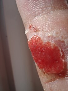
In medicine, granulation (from the Latin granulum = grain, grain ; granulation ) is the formation of young connective tissue that is visible to the naked eye in the course of wound healing. Granulation tissue is heavily permeated by capillaries (small blood vessels), which is why the surface appears "grainy" ( granulated ). Excess granulation tissue in skin wounds is referred to as caro luxurians ("wild meat") , as it can give the impression that it is growing wildly around the wound.
Process of granulation
Granulation occurs during the proliferative phase of the secondary healing on (with gaping wound edges and tissue defect). It begins after the exudation phase, about three to ten days after the injury. If there is no wound healing disorder, it will be over in about two weeks.
A fibrin network has formed through coagulation . Macrophages immigrated during the exudation phase activate various processes through their messenger substances: The growth factor β-FGF ( Fibroblast Growth Factor β), together with other messenger substances, ensures that fibroblasts migrate from the surrounding tissue. These lead to the synthesis of glycoproteins , proteoglycans and collagens . A kind of scaffold is created that is strengthened by the collagen fibers. New cells settle here. By angiogenesis adjacent, intact blood vessels get endothelial cells into the wound. These cells multiply and form new capillaries , which take care of the blood supply and thus the necessary exchange of substances . The resulting granulation tissue grows from the edge of the wound to the center until the defect is balanced and the wound is closed. Then new skin forms ( epithelialization ). The granulation tissue then undergoes some transformations, which end with the formation of solid scar tissue .
Cells of various types are also involved in the process of wound healing through granulation: fibroblasts ensure the closure of the tissue defect, angioblasts ensure the formation of new capillaries and keratinocytes ensure the epithelialization of the tissue. The messenger substances from the macrophages coordinate these processes.
Supportive wound treatment
Granulating wounds are covered with non-adhesive wound dressings, which protect the tissue when the dressing is changed and do not additionally injure the newly created capillaries . Overgrown granulation tissue can be treated (etched) with silver nitrate . If the growth is removed with the sharp spoon , healthy tissue can tear. In addition, there is a risk of pathogens being introduced into the wound, which cause an infection and thus impaired wound healing .
Laboratory medicine
In the laboratory medicine is referred to as granulation the microscopic and färbetechnischen detection of granules in cells . An example of a pathological finding is the toxic granulation of neutrophil granulocytes in the context of an acute bacterial infection.
Granulation is also a symptom of the blood count in promyelocytic leukemia .
literature
- Volker Schumpelick, Niels Bleese and Ulrich Mommsen (ed.): Short textbook of surgery . 8th edition. Georg Thieme Verlag, Stuttgart / New York 2010, ISBN 978-3-13-127128-0 , p. 24, 25, 38 .
- Susanne Danzer: wound assessment and wound treatment. Workbook for practice . 1st edition. Kohlhammer, Stuttgart 2012, ISBN 978-3-17-020671-7 .
