Bone segment navigation
With the help of bone segment navigation, missing bones are guided into the correct position in a computer-assisted surgical procedure and fixed by means of osteosynthesis . Bone segment navigation was first implemented in oral and maxillofacial surgery . Bone segments can be congenital or misaligned due to an accident. Such misalignments can affect the aesthetics and function of the organs in the mouth, jaw and face area; if the bony borders of the walls of the eye socket are affected, double vision can occur, if the temporomandibular joint is affected, this can lead to disruptions in the alignment of the teeth , if the entire roof of the skull is affected in terms of craniosynostosis , increased intracranial pressure can result.
Importance of surgical planning and simulation
The cutting of the bone and reattaching it in the correct position is called a conversion osteotomy . To ensure that the bone is really in the correct position after the operation, such a conversion osteotomy is planned and simulated in advance of the operation . This simulation is necessary to save operating time. Often, bone segments are not exposed at all or only to a small extent during the operation and remain covered by muscles, fatty tissue and skin - an assessment of whether a bone segment is in the correct position is difficult or impossible to make intraoperatively - this fact underlines the need for preoperative operation planning and simulation on an exposed bone model.
Aids for operation planning and simulation
Adjustment osteotomies as part of dysgnathic surgery are usually planned using plaster models of the jaw in an articulator . Adjustment osteotomies on the non-tooth-bearing bony skull can be planned on stereolithography models. These physical models have to be sawn up for the operation simulation and then reassembled. Since the 1990s, it has also been possible to simulate osteotomies on a computer directly on the preoperative CT or MRT image data set; this saves the high costs of creating the model and the time-consuming work of sawing and reassembling the physical models. The first system that allows such an operation simulation is the Laboratory Unit for Computer Assisted Surgery (LUCAS), which was developed at the University of Regensburg since 1998 with the support of Carl Zeiss Oberkochen.
Transfer of the surgical planning to the surgical site
The benefits of surgical planning depend on how well it is possible to reproduce the simulation of a conversion osteotomy on the patient. For the most part, the surgeon's sense of proportion was decisive for transferring the operation planning to the patient. In addition, various head frames were developed on which a bone segment offset was set with mechanical aids; The patient then had to wear such a head frame during the CT or MRT imaging; A further complicating factor was that the head frame had to remain unchanged in position from the CT or MRT imaging through the operation planning to the surgical procedure - this was relatively uncomfortable for the patient, and even completely impossible for children due to the lack of cooperation.

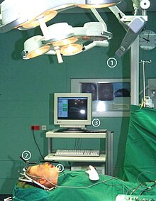
The first system that allowed bone segment navigation was the Surgical Segment Navigator (SSN), which was developed at the University of Regensburg since 1997 with the support of Carl Zeiss ( Oberkochen ). The system works without a head frame and consists of an infrared camera and infrared transmitters, which are anchored directly to the skull bone. At least three infrared transmitters are attached to the roof of the skull in order to record overall head movements. At least three other infrared transmitters are connected to the bone segment that is to be relocated as part of a repositioning osteotomy . The spatial position of the infrared transmitter - and thus also that of the bone segment - is measured by the infrared camera, the principle corresponds to that of satellite navigation . The workstation of the Surgical Segment Navigator (SSN) visualizes the starting and target positions of the bone segment to be moved and also marks the currently measured position of the loosened, freely movable bone segment. The bone segment is navigated according to the operation simulation, in that the visualization of the currently measured position is brought into congruence with the visualization for the target position.
Bone segment navigation is used to correct jaw misalignments in the context of dysgnathic surgery , for operative adjustment of the temporomandibular joint head, for reconstruction of the midface and the walls of the eye socket .
literature
- R. Marmulla: Computer Aided Bone Segment Navigation . Quintessenz, Berlin 2000, ISBN 3-87652-869-0 .
