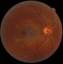
Yellow spot (eye)

The yellow spot , Latin macula lutea , macula or macula for short , is a narrowly circumscribed area in the rear, central area of the retina through which the visual axis runs and in the middle of which the distribution of color-sensitive sensory cells ( cones ) reaches its greatest density. The center of the macula forms the fovea centralis with the foveola ; the surrounding area is called the parafovea and, further peripheral, the perifovea . Depending on the perifoveal delimitation, the diameter of the yellow spot in adults is given as around 3 mm or around 5 mm.
The color of the area is caused, among other things, by pigments ( lutein and zeaxanthin ) embedded in the retina , but it is hardly noticeable in living people. The first anatomical description as a “yellow spot” comes from Samuel Thomas von Soemmerring , who relied on autopsy findings in humans.
About 15 ° nasal (towards the nose) of the macula lutea lies the almost 2 mm optic nerve papilla, which appears remarkably bright when the fundus is reflected , and is the cause of the blind spot in the visual field . There are no photoreceptor cells here, as this is where the inner nerve fibers of the ganglion cells of the retina leave the eye as an optic nerve , those from the macular region on the temple side. In addition, the central artery and the central vein enter and exit the eye here (see illustration).
Layout and function
The visual axis of the eye runs through the yellow spot , whereby the projected image usually falls on the funnel-shaped recessed central retinal area, which is also referred to as the “pit of vision” or fovea . Here the inner layers of the retina are shifted sideways, so that the sensory cells in the center - almost exclusively cone cells of the M and L types - can be reached by the incident light without being scattered by the overlying cell layers.
- In the area of the funnel-shaped deepened fovea centralis with a diameter of about 1.5 mm is the point for the highest visual acuity, because this is where the finest spatial resolution is possible. Particularly slim cones are located here in the closest packing, most of which are connected 1: 1 to the associated retinal ganglion cell. These smallest receptive fields are mainly found in the foveola (Latin for “dimple”), which is only 0.35 mm in diameter, in the middle of the fovea.
- In the approximately 0.5 mm narrow ring of the parafovea that surrounds it, there are increasingly more rods and the ratio of cones to rods is around 1: 1.
- The outermost edge of the macula is also called the perifovea . It contains considerably fewer cones and borders on the area of the highest rod density in the retina. If the perifoveal zone is not delimited as a narrow ring with an outer diameter of 3 mm, but an approximately 1.5 mm wide margin is assumed, the result is a total diameter of approximately 5.5 mm for the macula lutea .
Outside the macula, the incidence of cones in the peripheral retina decreases rapidly. In relation to the entire retina, the ratio of cones to rods is about 1:20 (6 million cones are compared to 120 million rods).
Through the eye movement, constantly changing areas of the environment are projected onto the fovea. The impression of a sharp overall picture arises in the brain instances downstream of the retina.
Diseases
- Macular degeneration
- Juvenile macular degeneration
- Vitelliform macular degeneration
- Retinopathia centralis serosa
- Macular ectopia
- Diabetic maculopathy
- Macular edema
- Macular telangiectasia
literature
- Theodor Axenfeld (founder), Hans Pau (ed.): Textbook and atlas of ophthalmology. With the collaboration of Rudolf Sachsenweger and others 12th, completely revised edition. Gustav Fischer, Stuttgart et al. 1980, ISBN 3-437-00255-4 .
- Herbert Kaufmann (Ed.): Strabismus. 3rd fundamentally revised and expanded edition. Georg Thieme, Stuttgart et al. 2004, ISBN 3-13-129723-9 .
Individual evidence
- ↑ a b Renate Lüllmann-Rauch: Pocket textbook histology. 3rd, completely revised edition. Georg Thieme, Stuttgart 2009, ISBN 978-3-13-129243-8 , p. 591.
- ↑ Web-Atlas Anatomie of the University of Mainz with electron microscopic images .
- ^ Franz Grehn: Ophthalmology. 30th, revised and updated edition. Springer, Heidelberg 2008, ISBN 978-3-540-75264-6 , p. 255.
- ^ Heinz Feneis (founder), Wolfgang Dauber: Feneis' Bild-Lexikon der Anatomie. 10th, completely revised edition. Georg Thieme, Stuttgart et al. 2008, ISBN 978-3-13-330110-7 .

