Magnetoencephalography
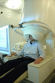
The magnetoencephalography (in English also magnetoencephalography , from Greek encephalon brain Graphein write), abbreviated MEG , is a measurement of the magnetic activity of the brain , performed by external sensors, so-called SQUIDs . The magnetic fields are usually first detected by coils or coil systems that are also superconducting and then measured by the SQUIDs. MEGs are complex and comparatively expensive devices. For the operation z. B. needs about 400 l of liquid helium per month for cooling.
Measurement of fields
The magnetic signals of the brain are only a few femto tesla (1 fT = T) and must be shielded as completely as possible from external disturbances. For this, the MEG is usually installed in an electromagnetic shielding cabin. The shielding chamber dampens the influence of low-frequency interference fields such as those caused by cars or elevators and protects against electromagnetic radiation. However, frequencies above one kilohertz ( Hz) have so far hardly been investigated with the MEG. Magnetic fields of external disturbances also differ from those of the brain in that their strength is much less dependent on location due to the greater distance to the place of origin. (The intensity decreases with the square of the distance.) With the help of the coil systems mentioned above, the fields with less spatial dependence can be suppressed very strongly. Therefore z. B. the heartbeat of the examined person with modern MEGs only a small interference effect. The earth's magnetic field is about 100 million times stronger than the fields recorded by the MEG, but it is very constant over time and only very slightly curved. Its influence is only disruptive when the entire MEG is exposed to mechanical vibrations.
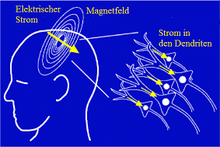
The magnetic signals of the brain are caused by the electrical currents of active nerve cells , which induce electrical voltages in the measuring coils of the MEG transducer . Therefore, in particular with the MEG, data can be recorded which express the current total activity of the brain without delay. Modern full-head MEGs have a helmet-like arrangement of up to 300 magnetic field sensors. A distinction is made between so-called magnetometers and gradiometers . Magnetometers have a simple pick-up coil. Nowadays gradiometers usually have two pick-up coils, which are arranged at a distance of 1.5 to 8 cm and wound in opposite directions. In this way, electromagnetic interference with little location dependency is suppressed even before the measurement. The very high time resolution (better than 1 ms ( second)), the ease of use of the high number of channels with precisely known sensor positions, as well as the numerically simpler modeling are the most important advantages of the MEG in the localization of brain activity compared to the EEG . Probably the biggest disadvantage of MEG localization is the ambiguity of the inverse problem . In short, it means that the localization can only be correct if the underlying model is essentially correct (number of centers and their rough local arrangement). This is where the advantages of metabolic functional methods such as fMRI , NIRS , PET or SPECT lie . By comparing and coupling the different functional methods, brain research provides ever more precise knowledge about the correct modeling of individual brain functions.
Newly developed mini sensors are able to carry out measurements at room temperature and measure field strengths of 1 picotesla. This opens up new design options and significant price reductions in the operation of the devices.
The MEG is a diagnostic method with good spatial and very high temporal resolution, which supplements other methods for measuring brain activity (functional methods), such as the EEG and the functional magnetic resonance method ( fMRI ). In medicine the MEG u. a. used to localize brain areas that trigger epileptic seizures or to perform complex cranial operations such. B. to plan in patients with brain tumors .
history
The first MEG was recorded by David Cohen at the Massachusetts Institute of Technology (MIT) in 1968. From 1985, a MEG was set up at the MEG Center Vienna by Lüder Deecke . It was the first generation with a 5-channel MEG system. In 1996 a MEG device with 143 channels followed (CTF Vancouver, Canada).
literature
- David Cohen: Magnetoencephalography: evidence of magnetic fields produced by alpha rhythm currents. In: Science. Volume 161, 1968, pp. 784-786.
- David Cohen: Magnetoencephalography: Detection of brain's electric activity with a superconducting magnetometer. In: Science. Volume 175, 1972, pp. 664-666.
- David Cohen: Boston and the history of biomagnetism. In: Neurology and Clinical Neurophysiology. Volume 30, 2004, p. 1.
- D. Cohen, E. Halgren: Magnetoencephalography. In: George Adelman, Barry H. Smith (Eds.): Encyclopedia of neuroscience. 3., corr. and exp. Edition. Elsevier Science, New York et al. a. 2004.
- M. Hämäläinen, R. Hari , R. Ilmoniemi, J. Knuutila, OV Lounasmaa: Magnetoencephalography - theory, instrumentation, and applications to noninvasive studies of signal processing in the human brain. In: Reviews of Modern Physics. Volume 65, 1993, pp. 413-497.
- W. Andrä, H. Nowak (Ed.): Magnetism in Medicine: A Handbook. 2nd Edition. Wiley-VCH, Weinheim 2006, ISBN 3-527-40558-5 . (English)
- Riitta Hari , Aina Puce: MEG-EEG PRIMER. Oxford Univ. Pr., Oxford 2017, ISBN 978-0-19-049777-4 .
Individual evidence
- ↑ NIST Mini-Sensor Measures Magnetic Activity in Human Brain (in English) http://www.nist.gov/pml/div688/brain-041912.cfm
Web links
- American Clinical MEG Society (English)
See also
- Computed tomography (CT)
- Positron Emission Tomography (PET)
- Magnetic resonance imaging (MRI)
- functional magnetic resonance imaging (fMRI)
- Electroencephalography (EEG)


