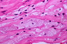
Foam cell
Foam cells form a large part of all known arteriosclerotically damaged blood vessels, which are also popularly known as “calcified”.
They arise in the intima and media layers of the vessel walls through fat deposits.
Foam cells are often observed in children and adolescents, but their formation there is usually only temporary (reversible). These yellowish gelatinous layers of fat, which medical professionals also refer to as fatty streaks , can cover 10 to 50% of the inner walls of the arteries. Investigations of these damages ( lesions ) showed that they are composed of both fat-laden macrophages and fat-laden smooth muscle cells . Tumorous accumulations of foam cells in the skin are called xanthomas .
The macrophages that normally circulate in the blood get into their preform, as monocytes , in the lower vascular layers by penetrating the endothelial cell layer . This process is stimulated by chemotactic factors. Only in the media (subendothelium) does a change in the appearance of the monocytes and a differentiation into macrophages take place. The macrophages can store lipids and cholesterol esters there. This in turn can lead directly to the formation of foam cells.
The observation that the lipid-laden macrophages can pass the endothelial cell layer and get back into the bloodstream triggered a controversy in medical research as to whether macrophages are useful (lipid removal) or harmful and are responsible for the development of arteriosclerosis, since they are released when they emerge the endothelial cell layer is injured. Some researchers suggest that the macrophages play a protective role in the early stages and a more harmful role later.
The mechanism of the formation of foam cells from smooth muscle cells and macrophages is only partially understood. There is agreement on the involvement of chemically modified (oxidized) LDL , which is absorbed and stored by the macrophages via scavenger receptors in an uninhibited manner and regardless of the concentration. These scavenger receptors are also found in smooth muscle cells. It can be assumed that mechanisms with which both types of cells could prevent an overload with lipids and cholesterol are inoperative or impaired.
