
Sialography
The sialography (from the Greek σιαλον, sialon, for saliva and γραφή, grafí, "the writing") is the representation of the ducts of the large salivary glands ( parotid , mandibular and sublingual salivary glands ). For this purpose, the respective mouth of the gland to be examined is probed with a fine, blunt cannula or catheter (the duct of the parotid gland is located in the cheek mucosa near the upper first molar ; the other ducts are located on the floor of the tongue behind the lower front teeth). Then a water-soluble contrast agent is carefully injected into the saliva duct under low pressure and x-rays of the gland and the ducts are made.
Sialography can be used, for example, in the diagnosis of salivary stones ( sialolithiasis ), in particular for localization. However, salivary stones are often visible on x-rays even without sialography. Furthermore, sialography can help diagnose salivary gland diseases.
In addition to sialography with conventional X-rays, there is also sialography with computed tomography and magnetic resonance tomography .

Sialography of the parotid gland in suspected Sjogren's syndrome
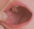
Opening of the parotid gland : parotid papilla

Opening of the mandibular salivary gland: Caruncula sublingualis


literature
- DM Yousem et al .: Major salivary gland imaging. In: Radiology , July 2000, 216 (1), pp. 19-29, PMID 10887223
- PM Som, HD Curtin: Head and Neck Imaging Fourth Edition, Mosby 2003, pp. 2013 ff., ISBN 0-323-00942-5
Individual evidence
- ↑ Mahmood F. Mafee, Galdino E. Valvassori, Minerva Becker: Imaging of the Head and the Neck. Thieme, 2005, ISBN 3-13-100942-X