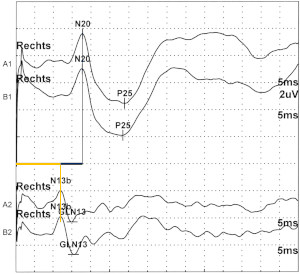
Somatosensitive evoked potentials
The somatosensitive evoked potentials (SEP or SSEP) are records of the electrical response of rapidly conducting sensory nerve fibers in the course of the loop path . After repeated electrical stimulation of a peripheral nerve at various points in the course of the procedure, mostly at the level of entry into the spinal cord and above the relevant brain area.
history
The SEP were first described by Dawson in 1947, at that time under difficult technical conditions, since electron tubes were available as amplifiers and oscillographs with documentation by photography were available for recording . The use of computer technology for averaging and recording has made things considerably easier.
principle
A sensitive nerve that is as close to the surface as possible is repeatedly stimulated electrically. As a result, an action potential spreads over the nerves, which can be derived with the help of surface electrodes (rarely also needle electrodes). Since the electrical voltages are very small, the potentials have to be freed from interference signals by means of averaging .
The derivation can take place in the course of the nerve (with the median nerve e.g. above Erb's point ), but mostly at the level of the entry into the spinal cord (“spinal potential” - with the median nerve e.g. above the spinous process of the 7th cervical vertebra) as well as over the corresponding area of the sensitive cerebral cortex ("cortical potential"). The difference between the spinal and cortical potential is called the “central lead time”. Due to the anatomy of the loop path, all three switchings (Nc.cuneatus / gracilis, thalamus, gyrus postcentralis) between the individual neurons occur in the central lead time.
Standard values
Relevant books contain standard values for the individual components of the SSEP. These should serve as a guide. Ultimately, every laboratory should develop its own standard values, since above all the amplitudes, but also the latencies of the potentials, depend on the technology used and the placement of the electrodes (not least the reference electrode).
Influencing factors
The length of the subject has a decisive influence on the result. Therefore, all standard values must be adapted to the body length. Body temperature also has an impact, especially cold extremities, on spinal latency. Age and posture also have less of an influence; there are contradicting information on gender.
application
The SSEP allow an assessment of the function of sensitive nerve tracts, initially in the course of the body, where they can hardly be examined by neurography due to the overlap with muscles, bones, etc. However, they are mainly used to test the central, sensitive path. If the myelin sheaths are damaged ("demyelination", e.g. multiple sclerosis ), the latency is longer; if the number of nerve fibers is reduced ("axonal damage") the amplitude of the potentials decreases.
The SSEP can also be used to estimate the prognosis in the event of severe brain damage (e.g. due to trauma or lack of oxygen ). The prognosis is very poor if a response from both sides can be derived from the spinal cord, but not from the cerebral cortex. This finding suggests bilateral brainstem damage that hardly allows an acceptable recovery. (see Firsching et al., Dtsch Arztebl 2003; 100 (27): A-1868)
literature
- K. Lowitzsch et al .: The EP book . Thieme 2000, ISBN 3131167319
- P. Vogel: Course book Clinical Neurophysiology . Thieme 2006, ISBN 313128112X

