Action potential
In physiology, action potential , abbreviated to AP , is a characteristic temporary deviation of the membrane potential of a cell from the resting potential . An action potential is formed automatically with cell-typical curve for a excitation (excitation) of the cell and spreads as an electrical signal through the cell membrane from. Colloquially, the action potentials of nerve cells are also called “nerve impulses”.
Only excitable cells can generate action potentials in response to stimuli or signals, through short-term changes in membrane conductivity as a result of interactions between special voltage-controlled ion channels in their membrane. Their time-dependent activation leads to different ion currents with correspondingly shifted potential differences. This results in an action potential curve in which the phase of repolarization follows the phase of depolarization after a possible plateau , with post-oscillating hyperpolarization . This process takes place automatically in a typical form when a certain threshold potential is exceeded and can only be triggered again after a certain refractory period .
In animals, excitable cells include not only nerve cells but also muscle cells and some secretory cells. Nerve cells receive stimuli or signals from other cells, convert them into membrane potential changes and can form action potentials (stimulation formation) as a cell-specific signal that is carried along the axon in a nerve fiber (→ excitation conduction ) and transmitted to other cells via synapses (→ excitation transmission ). Muscle cells are reached via neuromuscular synapses , which can also be excited and then form action potentials, which, guided by membrane protuberances, cause the muscle fibers to contract . Gland cells are reached via neuroglandular synapses ; special neuroendocrine cells can form action potentials, which are followed by the release of neurohormones .
Action potentials also occur in single cells - for example in paramecia and diatoms - as well as in multicellular algae ( candelabrum algae ), vascular plants ( mimosa ) and mushrooms .
history
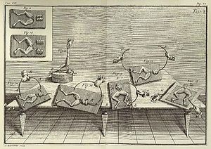
The Bolognese Luigi Aloisio Galvani discovered the phenomenon of muscle movements as a result of electrical forces as the twitching of the thighs of dissected frogs. His findings published in 1791 about an "animal electrical fluid" in nerves and muscles prompted Alessandro Volta to investigate galvanism , which led to the invention of the first battery in 1799 - called the Voltaic column . Volta also used this to study the effects of direct voltage on living beings.
In 1952, Alan Lloyd Hodgkin and Andrew Fielding Huxley presented a mathematical model that explains the formation of the action potential in the giant axon of the squid through the interplay of various ion channels and which became famous under the name of the Hodgkin-Huxley model . For this discovery, the two researchers, together with John Eccles, received the Nobel Prize in Medicine in 1963 .
Basics
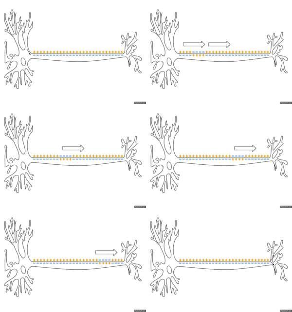
An action potential runs in a form typical for the cell type. With nerve cells it often only takes about one to two milliseconds, with skeletal muscle cells hardly longer, with heart muscle cells usually over 200 ms. There are no stronger or weaker action potentials depending on the stimulus, rather they are all-or-nothing answers. The signal strength therefore results from the frequency of action potentials. In nerve cells, they typically arise on the axon hill and are transmitted in series along the axon . Action potentials can also propagate backwards across the cell body and dendrites; the function of this forwarding is still being investigated. The axonal spread from the cell body to the terminal button is called orthodromic , the opposite is called antidromic .
The prerequisite for the development of an action potential are special properties of the cell's plasma membrane . The specific equipment with different groups of ion channels is reflected in the characteristics of the course form. Arousal occurs when the membrane potential moves away from the rest value and shifts towards less negative values. If this initial pre-depolarization reaches a certain threshold, the so-called threshold potential (around −55 mV), voltage-controlled ion channels are activated, which open in a chained sequence so that ion currents are enabled and then inactivated again.
During this chain of opening and closing processes of the channels, the membrane conductivities for different ions change temporarily. The associated short-term ion currents lead together to a characteristic potential curve. Its shape is the same for each cell, regardless of the strength of the triggering supra-threshold stimulus. The short-term changes in the potential now spread (electrotonically) to the neighboring membrane area and can then lead to the action potential again, which is the basis of the conduction of excitation.
Potential curve

Based on the resting membrane potential, which in neurons is between −90 and −70 mV depending on the cell type, four phases of the action potential are distinguished:
- In the initiation phase, a stimulus drives the negative voltage towards zero ( depolarization ). This can be done slowly or quickly and is reversible below the threshold potential. Such a stimulus can be a spatially approaching action potential or a post- synaptic ion current.
- If the threshold potential is exceeded, the depolarization accelerates strongly ( upstroke ). The membrane potential even becomes positive ( overshoot ).
- The maximum at +20 to +30 mV is followed by a return towards the resting potential ( repolarization ).
- In many neurons, the resting potential is initially undershot until z. B. −90 mV, and finally reached again from lower negative values. This is known as hyperpolarization or hyperpolarizing postpotential . No further action potential can be triggered during hyperpolarization.
The course of an action potential over time can extend over a few hundred milliseconds , for example in heart muscle cells. In the case of nerve cells, on the other hand, an action potential only lasts about 1–2 ms. After that, another action potential can be triggered, but not promptly, but only after a certain period of time. The cell is already in this so-called refractory phase during repolarization until it can again form an action potential under the same conditions.
A distinction is made here between the absolute refractory time (approx. 0.5 ms for neurons), in which no action potential can be triggered at all, and the relative refractory time (approx. 3.5 ms for neurons), in which stronger stimulus strengths are necessary because of the increased threshold potential are or only a deformed potential curve is to be triggered. The maximum frequency with which a neuron can form action potentials and transmit them in series as signals depends on the refractory periods.
causes
Understanding the action potential requires understanding the equilibrium potential for individual ions, as described in the article Membrane Potential. This voltage depends on the outside / inside concentration ratio and can be calculated using the Nernst equation . If only potassium channels are open, the Nernst potential of potassium (−90 mV) is established; if only sodium channels are open, the Nernst potential of sodium (+60 mV).
If the membrane is permeable to both potassium and sodium, the voltage is set at which the sum of the two currents is zero. The membrane potential is closer to the Nernst potential of an ion, the greater the permeability of the membrane for this ion; quantitatively this is described by the Goldman equation . At the resting membrane potential , it is mainly the potassium channels that are open, which explains the low voltage of around -70 mV. During an action potential, on the other hand, the permeability for sodium briefly predominates. All potentials that occur in the course of an action potential result from the permeabilities at the respective point in time.
The currents that occur in the course of an action potential are so small that they do not significantly change the concentrations on either side of the membrane. In order for the concentration ratios to remain constant in the long term, the work of the sodium-potassium pump is necessary, which uses ATP to remove three sodium ions in exchange for two potassium ions from the cell.
Properties of the ion channels
As described in the article on resting membrane potential , cells have a number of ion channels. Certain ion channels specific for sodium or potassium ions are primarily responsible for the animal action potential . These channels open depending on the membrane potential, so they are voltage-activated . At rest the membrane potential is negative.
For example, a voltage-dependent sodium channel (Na v channel) (also referred to as a fast sodium channel due to its property ) is closed and activated at the resting membrane potential . In the case of depolarization above a channel-specific value, the conformation of the transmembrane proteins changes. This makes the channel permeable to ions and changes to the open state . However, the channel does not remain open despite ongoing depolarization, but is closed again within a few milliseconds, regardless of the membrane potential. This usually happens through a part of the channel protein located in the cytoplasm, the inactivation domain, which is like a "plug" in the channel and clogs it. This state is called closed and inactivated .
The subsequent transition to the closed and activatable state is only possible after hyperpolarization (or complete repolarization in the case of heart muscle cells). The channel thus initially remains closed , can be activated after repolarization or hyperpolarization and can only then be opened again by depolarization. After opening, the canal only remains open for a short time, as it quickly changes into the closed form and is inactivated. However, a transition from inactivated to open is not possible with a depolarized membrane.
Not all channels open at the same time at the same value of membrane potential. Rather, the probability of a channel transitioning to a certain state is voltage-dependent. The purely statistical distribution results in an equilibrium such that a larger number of channels in total fulfills the model described above.
The time required to change from one state to the other is also channel-specific. In the sodium channel described, the conformational change from closed to open takes less than a millisecond, while a potassium channel takes around 10 ms for this.
Apart from the voltage, there are a number of other, often chemical, factors that cause the channels to open or close. For the action potential, only two of them are of a certain importance (see below). On the one hand, the inward rectifying potassium channels (K ir ) cannot be regulated per se. However, there are low molecular weight, positively charged substances such as spermine , which can clog the canal pores if there is sufficient depolarization (canal block, pore block). Another mechanism concerns potassium channels, which open when calcium ions bind to them intracellularly (usually intracellularly in very low concentrations).
procedure

Starting position
In the starting position the cell is at rest and shows its resting membrane potential. Almost all of the sodium channels are closed, only certain potassium channels are open. The potassium ions essentially determine the resting membrane potential. The direction and strength of all ion movements are determined by the electrochemical driving forces for the respective ions. Sodium ions in particular flow rapidly into the cell as soon as the channels open for this due to the prevailing concentration gradient .
Initiation phase
During the initiation phase, the membrane potential is changed in such a way that it deviates from the resting potential in the direction of negative values of decreasing magnitude until the reduction in the charge gradient reaches a certain threshold potential. This pre-depolarization can be done in the experiment by a stimulus electrode, on the axon mound by opening postsynaptic ion channels (Na + , Ca 2+ ) or on the axon membrane by an electrotonically transmitted (action) potential from a neighboring membrane region.
With such predepolarizing changes in the membrane potential, for example from −70 to −60 mV and beyond, K ir channels can be blocked by pore blockers such as spermine. This dampens a potassium current that has a rectifying effect in the direction of the resting potential. This makes it easier to reach the threshold potential and accelerates the subsequent depolarization when sodium channels are opened.
Spread and overshoot
At about −55 mV the voltage-gated sodium channels Na V begin to change into the open state. Sodium ions, which are far from their electrochemical equilibrium due to their high external concentration, flow in and depolarize the cell. This opens up further voltage-sensitive channels and even more ions can flow in: The fast spread leads to overshoot (polarization / charge reversal). The “explosive” depolarization after the threshold potential has been exceeded thus comes about through positive feedback .
Repolarization
Even before the maximum potential is reached in the overshoot, Na V channels begin to inactivate. At the same time, the voltage-dependent potassium channels K V come into play, K + ions flow out of the cell. Although these ion channels have their threshold at similar values, they take much longer to open. During the maximum of the Na conductivity these potassium channels are only half open; they reach their maximum when almost all Na channels are already inactivated. Therefore, the Na conductivity maximum is slightly before the voltage maximum in the overshoot, but the K conductivity maximum is in the phase of the steepest repolarization.
During the repolarization the potential approaches the resting potential again. The K V channels close and a block of pores in the K ir is broken, which is important for the stabilization of the resting potential. The Na V channels can slowly be activated again. Repolarization is also sometimes referred to as hyperpolarization, when this term is defined as the increasing negativity of a membrane potential.
Post hyperpolarization
In many cells, especially nerve cells, hyperpolarization beyond the resting potential can still be observed. It is explained by a continued increase in potassium conductivity, which means that the potential is even closer to the potassium equilibrium potential . The K-conductivity is increased because calcium ions flowing in during the action potential open special potassium channels here; it only normalizes when the intracellular calcium level drops again. If the repolarization has already been called hyperpolarization, this process of an additional lowering is then called post-hyperpolarization .
Refractory period
After the action potential has subsided, a cell cannot be excited for a short time . In the working myocardial cells of the heart, this phase - also called the "plateau phase" here - is particularly long-lasting, which is attributed to a "slow calcium influx". This fact is important because it prevents a retrograde re-entry of the excitation (unidirectionality). The duration of this period, the refractory period , depends on the time course of the reactivation of Na V channels. During the absolute refractory phase shortly after the overshoot, when repolarization is still in progress, these channels cannot reopen at all. It is also said that the threshold is infinite. During the relative refractory phase one needs stronger stimuli and receives weaker action potentials. Here the threshold value moves from infinity back to its normal value.
Threshold potential
Usually the triggering of an action potential is described as exceeding a certain threshold potential , from which sodium channels are opened in concert. Despite all efforts to find such an exact “fire threshold”, no fixed voltage value can be specified as a condition for an action potential. Instead, neurons fire on a relatively broad band of triggering membrane tensions. For this reason, neuroscientific refrains from the idea of a fixed value for the threshold potential. In terms of system theory, the process of creating an action potential can best be described by a bifurcation, such as in the Hodgkin-Huxley model . Nevertheless, it is quite common, also in the specialist literature, to continue to speak of a threshold in order to delimit the “gray area between calm and action potential”.
Animal action potentials
In Purkinje cells , the frequency of action potentials can be modulated not only by voltage-activated sodium channels but also by voltage-activated calcium channels .
Plant action potentials
In principle, cells of plants and fungi can also be excited electrically. The main difference to the animal action potential is that the depolarization does not occur through the influx of (positively charged) sodium ions, but through the outflow of (negatively charged) chloride ions . Together with the subsequent escape of (positively charged) potassium ions - which causes repolarization in both animal and plant cells - this means an osmotic loss of potassium chloride for plant cells ; on the other hand the animal action potential is osmotically neutral due to equal amounts of sodium influx and potassium outflow.
The coupling of electrical and osmotic events in the plant action potential suggests that electrical excitability in the common unicellular ancestors of animal and plant cells served to regulate the salt balance under variable salinity conditions , while the osmotically neutral transmission of signals by animal multicellular organisms with almost constant salinity is one represents evolutionarily younger achievement. Accordingly, the signaling function of action potentials in some vascular plants (for example Mimosa pudica ) has developed independently of that in animal cells.
literature
- Stefan Silbernagl, Agamemnon Despopoulos: Pocket Atlas of Physiology. 6th edition, Thieme Verlagsgruppe, Stuttgart 2003, ISBN 3-13-567706-0 .
Web links
- Action potential forwarding - Flash animation in 4 stages at informatik.uni-ulm.de
Individual evidence
- ↑ Machemer H, Ogura A: Ionic conductances of membranes in ciliated and deciliated Paramecium. . In: The Journal of Physiology . 296, 1979, pp. 49-60. PMID 529122 .
- ↑ Taylor AR: A fast Na + / Ca2 + -based action potential in a marine diatom . In: PLOS ONE . 4 (3), 2009, p. E4966. PMID 19305505 .
- ↑ a b Beilby MJ: Action potentials in charophytes . In: Int. Rev. Cytol. . 257, 2007, pp. 43-82. doi : 10.1016 / S0074-7696 (07) 57002-6 . PMID 17280895 .
- ↑ Sibaoka T: Excitable cells in Mimosa . In: Science . 137, 1962, p. 226. PMID 13912476 .
- ↑ a b Slayman CL, Long WS, Gradmann D: Action potentials in Neurospora crassa , a mycelial fungus . In: Biochimica et biophysica acta . 426, 1976, pp. 737-744. PMID 130926 .
- ^ A b Galvani: De viribus electricitatis in motu musculari ("On the forces of electricity in muscle movement"). Bologna, 1791; here online
- ↑ Piccolino M: Luigi Galvani and animal electricity: two centuries after the foundation of electrophysiology . In: Trends in Neuroscience . 20, No. 10, 1997, pp. 443-448. doi : 10.1016 / S0166-2236 (97) 01101-6 .
- ↑ Piccolino M: The bicentennial of the Voltaic battery (1800-2000): the artificial electric organ . In: Trends in Neuroscience . 23, No. 4, 2000, pp. 147-151. doi : 10.1016 / S0166-2236 (99) 01544-1 .
- ↑ Hodgkin AL, Huxley AF: A quantitative description of membrane current and its application to conduction and excitation in nerve . In: J. Physiol. . 117, 1952, pp. 500-544. PMID 12991237 .
- ^ John PJ Pinel, Paul Pauli: Biopsychology. Pearson Studies; Edition: 6th, updated. Edition (May 29, 2007), ISBN 3-8273-7217-8 , p. 110.
- ↑ Robert F. Schmidt : Physiology of humans: With pathophysiology. Springer Verlag, 2007, ISBN 978-3-540-32908-4 , p. 88.
- ↑ E. Hosy, C. Piochon, E. Teuling, L. Rinaldo, C. Hansel: SK2 channel expression and function in cerebellar Purkinje cells. In: The Journal of Physiology. Volume 589, Pt 14 July 2011, pp. 3433-3440, ISSN 1469-7793 . doi: 10.1113 / jphysiol.2011.205823 . PMID 21521760 . PMC 3167108 (free full text).
- ↑ N. Zheng, IM Raman: Synaptic inhibition, excitation, and plasticity in neurons of the cerebellar nuclei. In: Cerebellum. Volume 9, Number 1, March 2010, pp. 56-66, ISSN 1473-4230 . doi: 10.1007 / s12311-009-0140-6 . PMID 19847585 . PMC 2841711 (free full text).
- ↑ Mummert H, Gradmann D: Action potentials in Acetabularia : measurement and simulation of voltage-gated fluxes . In: Journal of Membrane Biology . 124, 1991, pp. 265-273. PMID 1664861 .
- ^ Gradmann D: Models for oscillations in plants . In: Austr. J. Plant Physiol. . 28, 2001, pp. 577-590.
- ^ Gradmann D, Hoffstadt J: Electrocoupling of ion transporters in plants: Interaction with internal ion concentrations . In: Journal of Membrane Biology . 166, 1998, pp. 51-59. PMID 9784585 .
- ^ D. Gradmann, H. Mummert: Plant action potentials. In: RM Spanswick, WJ Lucas, J. Dainty: Plant Membrane Transport: Current Conceptual Issues. Elsevier Biomedical Press, Amsterdam 1980, ISBN 0-444-80192-8 , pp. 333-344.

