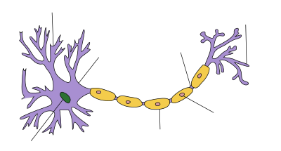
Axon
| Structure of a nerve cell |
|---|
The Axon , rarely the axon (by Alt Gr. Ὁ ἄξων ho axon , axis'), and axon or axis cylinder mentioned, is an often long tube-like nerve cell process, a neurite , in a case of glial cells runs and together with the latter serving as a nerve fiber designated becomes. Lateral branches of the axon are also called its collaterals and, like the terminal axon, can branch into several terminal boxes.
Most neurons have a single axon. But there are also nerve cells that do not have an axon, e.g. B. Different amacrine cells of the retina.
While an axon transmits an impulse from the cell, dendrites receive signals from other cells.
Anatomy of the axon
An axon is the extension of a nerve cell surrounded by a glial envelope, via which signals are directed, mostly in the form of action potentials . In the course of this, the following sections can be distinguished:
- The cone of origin or axon mound emerges as a pyramidal protrusion directly from the perikaryon; this area, free of Nissl substance, marks the beginning of a nerve cell extension that becomes an axon.
- The initial segment is the subsequent short segment of the extension and always without a cover. Since the excitation threshold of the axolemma is extremely low in the initial segment, an action potential can easily be initiated here, which is passed on as excitation.
- The main course of an axon can be of different lengths and also have branches along the way, which are referred to as collaterals .
- As an end branch , an axon sometimes branches off like a tree to form a telodendron . A nerve cell can be connected to several other nerve cells or effectors through this end tree. The telodendria, which can also occur in collaterals, end in a large number of end sections as presynaptic endings , also called axon terminals , end knobs or boutons , each of which represents the presynaptic part of a synapse .
The cytoplasm of an axon is called axoplasm and differs in some ways from that of the nerve cell body (perikaryon or soma). Mitochondria and vesicles are also found in the axon, but only in exceptional cases ribosomes or a rough endoplasmic reticulum. The maintenance and function of the axon are therefore dependent on protein synthesis in the cell body. When severed, the severed cell process dies.
Both neurites and dendrites contain a cytoskeleton made up of neurofilaments and neurotubules . In most cases, the neurite transmitting signals from the cell body is encased by glia to the axon. In this case, an axonal microtubule differs from a dendritic one on the one hand by the associated proteins and on the other hand by its orientation: the axonal microtubules are then oriented with their plus end (the growing end) towards the axon end. In the case of the dendritic, the plus end can also be in the cell body.
In humans, there are axons less than a millimeter long and over a meter long, such as the processes of motor nerve cells in the spinal cord that innervate the toe muscles. The diameter of an axon conducting action potentials can be between 0.08 µm and 20 µm and remains relatively constant over the entire length. The finest neurites with about 0.05 µm were found in amacrine cells in the lamina of the visual system of fruit flies and conduct graduated changes in potential as signals. The giant axons of squids are among the thickest axons with a diameter of up to one millimeter (1000 µm) ; they innervate the muscles surrounding the sipho , the contraction of which allows rapid escape movements due to water recoil. These axons conduct action potentials, but are not surrounded by a myelin sheath in such a way that saltatory conduction is possible.
Such an axon from an octopus is 50 to 1000 times thicker than one from mammals . Due to the larger axon cross-section, the longitudinal resistance of the axon (internal resistance) is significantly lower, so that the electrotonic current flow from the excited to the still unexcited areas can take place more easily, which enables faster transmission. However, despite the large axon diameter , the speed of the conduction of excitation is still lower than with axons that are a hundred times thinner when these are myelinated.
Myelination
A myelin sheath around axons is formed by the glial cells of the nervous system, in the central nervous system (CNS) by the oligodendrocytes and in the peripheral nervous system (PNS) by the Schwann cells . With myelination, a different, abrupt transmission of electrical signals via the axon becomes possible, which requires significantly less energy, allows a thinner axon (space and material savings) and is also faster than continuous transmission .
The unit consisting of the axon of a nerve cell and the envelope structures of glial cells, including the reinforced basal lamina in peripheral nerves , is called nerve fiber . Nerve fibers in which glial cells have wrapped themselves several times around the axial cylinder, so that a myelin-rich sheath is formed, are called myelinated or medullary . According to the thickness of the myelin sheath or myelin sheath, a distinction is made between rich and poor fibers; unmyelinated or not myelinisert are nerve fibers without designed as a myelin sheath wrapping, z. B. when axons run in simple folds of glial cells.
Only with the formation of a myelin sheath can an axon be electrically isolated from its environment in such a way that signals can be passed electrotonically quickly over longer sections without significant attenuation and only refreshed again in the gaps between successive glial cells - on the so-called Ranvier cord rings Need to become. This abrupt (saltatory) forwarding of action potentials makes significantly higher conduction speeds possible; for example, it is six times faster at one fiftieth the diameter of giant axons.
The thickness of the medullary sheath depends on the number of windings on the part of the glial cell and is fully developed depending on the axon diameter. Thicker axons have thicker myelin sheaths with up to around a hundred lamellar layers. The width of the Ranvier cord rings or knots varies little; the distance between these knots, the internode , can be between 0.1 mm and 1.5 mm. The length of an internode corresponds to one Schwann cell in a peripheral nerve. Since their number along a nerve fiber does not change later as the body grows, the internode length increases with growth. For larger diameter of myelinated axons, the rate of conduction is also higher in myelinated nerve fibers, so that a division of nerve fibers alone after the conduction velocity usually about also delivers to the fiber thickness (see classification according to line speed by Erlanger / Gasser and nerve conduction velocity ).
Myelinated axons are modified to match the type of conduction. In the course of the axon, the axolemma, the cell membrane of a nerve cell then alternates between short sections with denser ion channels in the area of the Ranvier nodes ( nodal ) and longer sections with sparser populations ( internodal axolemma ).
Growth and development
Axon growth begins directly with aggregation. Both growing axons and dendrites have a growth cone with finger-like extensions (filopodia). These offshoots “grope” for the way.
This hypothesis is based on chemotrophic factors that are emitted by the target cells. The phenomenon was first detected in the optic nerve (optic nerve) of the frog.
This is based on signals that are emitted by axons and ensures that regrowing axons have an affinity for the same path.
tasks
The axon conducts electrical nerve impulses away from the cell body ( perikaryon or soma ). However, the transmission from nerve cell to nerve cell or to the successor organ is usually not electrical, but chemical. At the end button , neurotransmitters are released as chemical messenger substances that bind to a receptor , thereby influencing the membrane permeability for certain ions and thus causing a voltage change in the associated membrane region of the downstream cell.
Depending on the direction of the conduction of excitation, a distinction is made between afferent and efferent axons. In relation to the nervous system as a whole, afferent neurites conduct excitation from the sense organs to the CNS. These afferents are divided into somatic (from the body surface) and visceral (from the intestines). Efferent neurites, on the other hand, conduct impulses from the CNS to the peripheral effectors (e.g. muscles or glands); Here, too, a distinction is made between somatic (from motor neurons to skeletal muscles, e.g. of the foot) and visceral efferents (for smooth muscles and heart muscles as well as glands).
Axonal transport
In addition to the transmission of electrical signals, substances are also transported in the axon. A distinction is made between slow axonal transport, which only runs in one direction, from the cell body (soma) to the peripheral end of the axon, and fast axonal transport, which takes place in both directions - both anterograde and retrograde, from the terminal axon to the soma.
history
After it was recognized that nerves, despite their similar appearance, are not tendons that connect muscles and bones ( old Greek νεῦρον neuron 'flechse, tendon'), but rather form a connection that runs through the entire body, various models were developed for their tasks. So also mechanistic ones like that of René Descartes 1632 in his "Treatise on man" ( Traité de l'homme ; posthumously De homine 1662), according to which their fibers would be able to produce movements by means of mechanical pull, similar to a machine . The light microscopes , which were further developed in the 17th century, allowed increasingly finer insights into the structure of tissue, and the discovery of galvanic currents towards the end of the 18th century made other ideas about how it worked.
But studies with intracellular recordings from individual neurons in the nervous system could only be carried out by K. Cole and H. Curtis in the 1930s. Prior to this, peripheral nerves were examined, the nerve fibers bundled within them were examined more closely and their course traced. In 1860 the German anatomist Otto Deiters was already familiar with "the transition from an axis cylinder of genuine nature to a ganglion cell process"; He is credited with being the first to distinguish the only “main cell process” from other “protoplasmic processes”, for which the Swiss anatomist Wilhelm His later coined the term “dendrites”. The Swiss Albert von Kölliker and the German Robert Remak were the first to identify and describe the initial segment of the axon.
At the giant axon of squid of was Alan Lloyd Hodgkin and Andrew Fielding Huxley formation and conduction of action potentials investigated and 1952 as Hodgkin-Huxley model quantitatively described.
Illnesses and injuries
Axotomy and degeneration
An axotomy is the severing of an axon. This can happen as a result of an accident or is part of controlled animal experiments. The controlled transection of axons led to the identification of two types of neuronal degeneration (see also neuronal plasticity , apoptosis , necrosis ).
- Anterograde degeneration
This degeneration of the cut off far (distal) part of the affected neuron, i.e. the terminal axon and some collaterals, occurs quickly because the distal part is dependent on the metabolic supply from the soma.
- Retrograde degeneration
If the severed site is close to the cell body, degeneration of the near (proximal) segment can also occur. This proceeds more slowly and manifests itself after two to three days through degenerative or regenerative changes in the neuron. The course depends crucially on whether the neuron can resume synaptic contact with a target cell.
In the worst case, neighboring neurons can also degenerate. Depending on the location of the additionally affected neurons, one speaks of anterograde or retrograde transneural degeneration .
regeneration
The original ability to precisely grow axons during the development of the nervous system is lost in the mature human brain. Neuroregeneration does not usually take place in the CNS. Dead neurons are replaced by glial cells (mostly astrocytes) and so-called glian scars develop .
Neuroregeneration in the PNS usually begins two to three days after the axon has been injured and depends largely on the type of injury to the neuron:
- If the myelin sheaths are still intact (for example after being squeezed), the axon can grow back in them to their original destination at a rate of around 2-3 mm per day (complete functional regeneration).
- If the severed ends are still close to each other, regrowth in the myelin sheaths is also possible, but then often to the wrong destination (difficult functional regeneration)
- If the severed ends are far apart or if there is extensive damage, functional regeneration is in most cases not possible without surgical interventions and, even after this, in many cases only incomplete.
Demyelinating diseases
Demyelinating diseases (demyelinating diseases) cause the axons in the CNS to lose parts of their myelin sheath and thus parts of the myelin sheath are destroyed. This is e.g. This is the case, for example, with multiple sclerosis (MS), Baló's disease , acute disseminated encephalomyelitis (ADEM) or neuromyelitis optica (Devic's syndrome).
Web links
- Entry to Axon in Flexikon , a Wiki of the DocCheck company
- Axon - Article at Wissenschaft-Online .de
Individual evidence
- ↑ axon. In: Lexicon of Neuroscience. Retrieved November 27, 2015 . - Designations at Spektrum.de .
- ↑ a b c Clemens Cherry: Biopsychologie from A to Z . Springer textbook, ISBN 3540396039 , p. 30/31 Lemma "Axon (afferent / efferent)".
- ^ A b c Luiz Carlos Junqueira (author), José Carneiro (author), Manfred Gratzl (editor): Histology: New Approval Regulations . Springer, Berlin; Edition: 6th, newly translated. revised A. (September 15, 2004). ISBN 354021965X , page 112/113.
- ^ Theodor H. Schiebler, Horst-W. Korf: Anatomy: histology, history of development, macroscopic and microscopic anatomy, topography . Steinkopff; Edition: 10th, completely revised. Edition (September 21, 2007), ISBN 3798517703 , page 72.
- ^ Aldo Faisal et al .: Ion-Channel Noise Places Limits on the Miniaturization of the Brain's Wiring . In: CurrentBiology , Volume 15, Issue 12, June 2005, p. 1147 .
- ↑ Renate Lüllmann-Rauch: Pocket textbook histology. 3. Edition. Thieme Verlag, 2009, ISBN 978-3-13-129243-8 , p. 191.
- ↑ Clemens Cherry: Biopsychologie from A to Z . Springer textbook, ISBN 3540396039 , p. 49 Lemma "Chemoaffinity hypothesis".
- ↑ Otto Deiters (Max Schultze (ed.)): Studies of brain and spinal cord of humans and mammals , Vieweg, Braunschweig 1865, page 2 .
- ^ A b John PJ Pinel, Paul Pauli: Biopsychology , PEARSON STUDIUM; Edition: 6th, updated. Edition (May 29, 2007), p. 327.
