Schwann cell
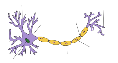
| Structure of a nerve cell |
|---|

The Schwann cell ( Gliocytus periphericus , also Schwann cell ) is a special form of a glial cell . It forms a sheath and support cell that surrounds the axon of a nerve cell in the peripheral course and electrically insulates myelin sheath in myelinated fibers . The lemnocyte adjacent to the ganglion cells is also viewed as a Schwann cell in the broader sense.
history
It was named after its discoverer, the German anatomist and physiologist Theodor Schwann (1810–1882).
Layout and function
Schwann cells only exist in the peripheral nervous system . They are used to form myelin sheaths , which isolate the axons so that their conduction speed is increased. In the central nervous system , oligodendrocytes perform the same function .
Schwann cells attach to axons of nerve fibers. At first it remains undetermined how many nerve fibers and to what extent they do this. If they are only partially or simply attached to the axons, they are still referred to as medullary nerve fibers. Only the fat-protein mixture created by multiple wrapping is referred to as myelin or the nerve fiber as myelinated . In contrast to the oligodendrocytes, the Schwann cells are not able to completely encase several axons; should an attachment to several axons take place, this will never lead to a complete wrapping. Another difference is that Schwann cells can induce axonal regeneration, or regrowth of the axon, after injury - an ability that oligodendrocytes do not have, which is why regeneration of the CNS is limited in mammals.
In about a third of all nerve fibers, the Schwann cell wraps itself around an axon several times during growth. Only in vertebrates can the Schwann cell wrap itself around an axon in many ways.
The cytoplasm and the cell nucleus are located in the outer area of a Schwann cell, which does not wrap itself around the axon and thus does not participate in the myelin . This part is also called the Schwann sheath or neurolemm . It is surrounded by a basal lamina that connects it to the surrounding connective tissue endoneurium . When an axon grows back after injury, the survival of the basal lamina is important because it guides the axon.
There are regular interruptions in the myelin sheath between neighboring Schwann cells along the axon. These are called Ranvier tie rings . This is where the saltatory excitation conduction, which is important for increasing the speed of nerve conduction, takes place . The section between two Ranvier rings is called the internode .
Since vertebrates have Schwann cells, but other species, e.g. B. arthropods , this is an evolutionary advantage for large vertebrates due to the significantly higher reaction speed. This advantage disappears in smaller animals, since the chemical transmission of the information in the synapses takes up most of the time.
Diseases
Acquired inflammatory diseases that involve Schwann cells include chronic inflammatory demyelinating polyneuropathy (CIDP), Guillain-Barré syndrome (GBS) and leprosy . Among the hereditary diseases with Schwann cell involvement are among the Charcot-Marie-Tooth disease , the Krabbe disease and Niemann-Pick disease . Benign tumors of the Schwann cells are called schwannomas or neuromas. Multiple sclerosis (MS) as a result of the autoimmune, inflammation-initiated destruction of the myelin sheath can also be mentioned here. The efforts of medical research aim at repairing and breaking the chain of origin. There are extensive statistics on serial medication and the like. a. with cortisone followed by vector observation.
Individual evidence
- ↑ Clemens Cherry: Biopsychologie from A to Z . Springer textbook, ISBN 3-540-39603-9 , p. 254 Lemma "Schwann cells".
- ↑ Thomas Heinzeller, Carl M. Büsing: Histology, Histopathology and Cytology for an introduction . Georg Thieme, Stuttgart 2001, ISBN 978-3-13-126831-0 , p. 66 .
- ^ John PJ Pinel, Paul Pauli: Biopsychology , PEARSON STUDIUM; Edition: 6th, updated. Edition (May 29, 2007), ISBN 3-8273-7217-8 , p. 78.
- ^ Niels Birbaumer, Robert F. Schmidt: Biological Psychology (Springer textbook), Springer Berlin Heidelberg; 7., completely revised. u. supplemented edition (July 21, 2010), ISBN 3-540-95937-8 , p. 26.
- ↑ Ulrich Welsch : Sobotta textbook histology. Cytology, histology, microscopic anatomy . 2nd, completely revised edition. Elsevier, Urban & Fischer, Munich et al. 2006, ISBN 3-437-44430-1 , pp. 187 .
- ↑ Clemens Cherry: Biopsychologie from A to Z . Springer textbook, ISBN 3-540-39603-9 , p. 236 Lemma "Ranviersche Schnürringe".

