Light microscope
Light microscopes (from the Greek μικρόν ( micrón ) = "small", and σκοπεῖν ( skopein ) = "to look at something") are microscopes that generate greatly enlarged images of small structures or objects with the help of light . The magnification takes place in accordance with the laws of optics using light refraction on glass lenses .
In order to be able to recognize structures in the generated image, the image must contain sufficient contrast that is present in many biological objects such as B. tissue sections or small aquatic life is hardly present. The 'typical' microscopic procedure for such objects is bright field microscopy , in which the contrast is created by colored or dark structures in the illuminated specimen, reinforced if necessary by additional artificial coloring of the object. In the case of colorless specimens, contrast can also be created using special lighting methods by converting differences in optical density (refractive index) into differences in brightness. This happens with dark field microscopy , phase contrast microscopy and with differential interference contrast (DIC) or with the method with oblique illumination, which was already used in the early days of microscopy. Differences in the polarization behavior of the specimen are used in polarization microscopy . Fluorescent structures in the specimen are a prerequisite for fluorescence microscopy and its numerous special procedures. Other microscopic methods are confocal microscopy and multiphoton microscopy . All of these procedures are covered in their own articles. The article here presents common principles of various microscopic procedures.
How simple and compound microscopes work
Light microscopy can be performed with “simple” or “compound” microscopes. Today's microscopes are typically “compound microscopes”.
Simple microscopes
Simple microscopes only have a single optical system for magnification and work like a magnifying glass (for the principle of magnification, see there). Originally only a single glass lens was used for this . A very short focal length is required to achieve the high magnification typical of microscopes. The resulting strong curvature of the lens surface means that the lens must have a small diameter in the millimeter range. It must be held close to the eye at a correspondingly small distance, which is exhausting and has led to the general lack of use of these microscopes. In the simplest case, a simple microscope only consisted of a glass lens and a holder for it.
The best known are probably those devices built by Antonie van Leeuwenhoek , with which he made numerous scientific discoveries at the end of the 17th century. The replica of such a microscope shown in the figure is held with the side lying on the surface close to the eye. On the right of the picture, at the end of a rhombus, you can see a point on which the specimen was mounted. Below that the glass lens is embedded in the metal plate.
In the course of time, numerous variants of simple microscopes have been developed, such as B. the "flea glass", the compass microscope, simple "screw-barrel" microscopes, classic dissecting microscopes and the so-called botanical microscopes. In the search for better imaging quality, gemstone lenses were also used in the 19th century because of their high refractive index and thus lower spherical aberration, or a combination of two or three planoconvex lenses (doublet, triplet) also to reduce imaging errors. There were also simple microscopes with combinable single lenses with screw mounts to change the magnification.Such simple microscopes were offered until the end of the 19th century. For example, after the company was founded in Jena, from the 1850s onwards, Zeiss produced doublets up to 125 times magnification and triplets up to 300 times magnification, and in 1895 another doublet 70 times.
Since a simple microscope with high magnification must be held close to the eye, the specimen can usually only be illuminated from the back. As a rule, transmitted light is used. However, since Johann Lieberkühn (1740) invented a concave illuminating mirror that surrounds the lens and is directed towards the object, there has also been the possibility of incident light illumination. The simple "natural scientist microscopes" with lower magnification were mostly reflected light microscopes.
Two-stage magnification with the compound microscope
Combined microscopes consist of at least two optical systems connected in series, each with its own magnification. The front lens , the objective , creates an enlarged real image , the intermediate image , which is enlarged a second time by the eyepiece . The eyepiece works like a magnifying glass and creates a virtual image of the intermediate image . The total magnification of the microscope is the product of the objective magnification and the eyepiece magnification. With a 20x objective and a 10x eyepiece, the total magnification is 200x.
The first compound microscopes consisted of only two individual lenses, and very soon the eyepiece was made up of two lenses to enlarge the usable image field and reduce aberrations (e.g. Huygens eyepiece ). In modern microscopes, objectives and eyepieces consist of several lenses to compensate for various optical aberrations . Here is about the chromatic aberration to name who could be limited only in the 19th century by introducing new types of glass. Since the aberrations of the objective and eyepiece are multiplied, compound microscopes were initially inferior to simple microscopes. The objectives and eyepieces are usually interchangeable so that the magnification can be adapted to the task at hand.
Composite microscope designs
Transmitted light or reflected light microscopy
Robert Hooke worked with a reflected light microscope in the 17th century.
Depending on the side from which the light falls on the specimen, a distinction is made between incident and transmitted light or between incident and transmitted light microscopy.
In transmitted light microscopy , the illumination is passed through the specimen from behind before it is picked up by the objective of the microscope (orange arrows in the diagram). Therefore, transparent or thinly sliced preparations are required. This technique is used in the most common microscopic method, transmitted light bright field microscopy .
With reflected light microscopy , the light is either directed from the microscope through the objective onto the specimen (light blue arrows) or irradiated from the side (green arrow). The light reflected by the specimen is in turn captured by the objective. Incident light microscopy is also possible with opaque specimens. Such preparations are common in materials science, for example, where specimens of a material are ground and polished or etched surfaces, which are then examined microscopically. Incident light illumination through the objective is also widespread in fluorescence microscopy . Stereo microscopes usually work with lateral incident light.
Lateral illumination (magenta-colored arrow) was or is used in some special procedures (see slit ultrasonic microscope and light disk microscopy ).
Structure of a typical compound transmitted light microscope
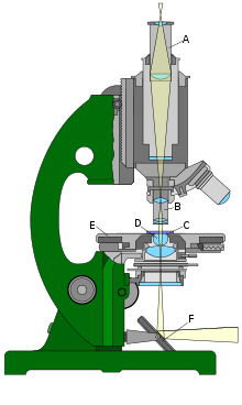
The components of a typical transmitted light microscope work together as follows:
- The lens (B) creates a real image , the intermediate image . Modern microscopes are usually equipped with several objectives that are mounted in an objective nosepiece. This enables the objective to be changed quickly by turning the turret.
- The intermediate image is enlarged one more time by the eyepiece (A). The intermediate image plane typically lies within the eyepiece. The total magnification of the microscope is calculated by multiplying the magnifications of the objective and eyepiece. Many eyepieces have a 10x (10 ×) magnification. Frequent objective magnifications are between 10 × and 100 ×.
- The tube between the objective and the eyepiece is called the tube .
- The preparation (also: object) is usually attached to the glass slide (C) in transmitted light microscopes . The slide is attached to the specimen table (E).
- To ensure that the light coming from below optimally illuminates the object, transmitted light microscopes have a separate lens system, the condenser (D). This is attached to the stage.
- The stage can be moved up and down to focus on the object. The condenser is moved along with it.
- A mirror (F) serves as a light source for old and very simple new microscopes. Otherwise an electric light source is used.
The preparation can be illuminated by means of critical lighting or Köhler lighting (see below).
Tube length, finite optics and infinite optics
The objectives of older or smaller microscopes are adapted to a defined tube length and generate a real intermediate image at a precisely defined distance, which is then enlarged by the ocular optics. The manufacturers agreed on a tube length of 160 mm, with older microscopes this tube length may differ. The Leitz / Wetzlar company manufactured according to an in-house standard of 170 mm.
However, this defined tube length has some disadvantages. Optical elements and assemblies cannot simply be inserted into the beam path, since z. B. simply there was not enough space for this. Newer microscopes are therefore equipped with so-called “infinite optics”. In this case, the lens does not create a real intermediate image, but the light leaves the lens as infinite parallel rays, which enables an "infinitely" long tube. Any number of intermediate elements such as filters, beam splitters, etc. can thus be inserted into the beam path. Since no image can emerge from the parallel rays of light, there is a tube lens at the end of the infinite tubes. This creates a real intermediate image from the parallel light beams, which can then be enlarged again by the ocular optics. Infinite optical lenses can usually be recognized by the ∞ ( infinity symbol ) on them.
In contrast to the infinite optics, the classic optics with a fixed tube length is called "finite optics". The length of the intended tube is indicated in millimeters on appropriate lenses, around 160 or 170.
Upright and inverted (also: inverted) microscopes
A microscope with the objective above the specimen is called an upright microscope. With transmitted light microscopes, the light then comes to the specimen from below. Above is the lens through which the light goes up to the eyepiece. This is the more common type.
If this light path is reversed, one speaks of an inverted or inverted microscope. With transmitted light illumination, the light falls onto the specimen from above, and the objective is located below it. In order to enable comfortable work, the light is then deflected so that you can look into the eyepieces from above (see illustration).
Inverted microscopes, for example, for observation of the cell culture - cells employed, since stopping the cells at the bottom of the culture vessel. The distance from the cells to the objective would be too great with an upright microscope. Microscopes with this design are an indispensable instrument for examinations of living cells in culture vessels ( cell culture ), e.g. B. in the patch-clamp technique and when using micromanipulators that are brought up to the preparation.
Illumination of the preparation
There are two common lighting methods to illuminate the field of view brightly. The critical lighting is historically older. It is still used today in some very simple microscopes. The Köhler lighting developed by August Köhler allows the preparation to be illuminated more evenly. Today it is standard in routine and research microscopes. Transmitted light bright field microscopy with Koehler illumination is typically the starting point for the application of special light microscopic contrast methods such as phase contrast and differential interference contrast . Both lighting methods were originally developed for transmitted light brightfield microscopy, but are also used in other methods, such as fluorescence microscopy.
Critical lighting
With critical lighting, a reduced image of the light source is generated in the preparation plane. If a light bulb is used as the light source, the filament is imaged in the plane of focus with the help of the condenser . This ensures that the specimen is illuminated with the maximum possible brightness. The focal length of a microscope condenser is usually quite short. In order to be able to generate an image of the light source in the focal plane of the microscope, the condenser must first be positioned close to the specimen. Second, the light source must be comparatively far away from the condenser so that it is clearly in front of its front focal plane . In order to prevent the image of the filament from making it difficult to identify the specimen structures, a frosted glass filter is placed in the illumination beam path below the condenser. If this is not sufficient, the condenser can be lowered a little so that the image of the filament becomes blurred.
If a daylight mirror is not used for lighting, this is usually flat on one side and hollow on the other. The concave mirror can be used for objectives with low magnification when the condenser has been removed. At higher magnifications, the illumination must be condensed onto a smaller area of the specimen. This is done with the condenser using the plane mirror . With daylight lighting, structures from the environment such as window frames can be shown in critical lighting. A frosted glass filter under the condenser or a condenser lowering can also help here.
Köhler lighting
At the end of the 19th century August Köhler dealt with microphotography , that is, photography with the help of a microscope. When observed directly through the eyepiece , the uneven brightness of the field of view was comparatively less disturbing in critical lighting, since the specimen could be moved back and forth as required. In photomicrography, however, uneven lighting resulted in poor image quality. He therefore developed a process that allowed uniform brightness with the same high overall brightness. He published this method, which is named after him today, in 1893. The process of setting the Koehler illumination is called Koehler.
In addition to uniform field of view illumination, Köhler lighting has another advantage: only the area of the specimen that is actually being observed is illuminated. This avoids disruptive stray light that would arise in neighboring areas. With this type of illumination, an image of the light source is not generated in the specimen plane, but in the plane of the diaphragm below the condenser. This is called the condenser diaphragm or aperture diaphragm. In contrast, an image of the luminous field diaphragm (also: field diaphragm) is sharply imaged in the preparation plane. This aperture is located near the light source, usually it is built into the base of the microscope. The image in the specimen plane is brought into focus by moving the condenser up or down. Köhler lighting is only possible with an artificial light source.
A Kohler microscope has two interrelated, intertwined beam paths and each of the two has several 'conjugate planes', that is, what is in focus in one of the planes is also in focus in the other conjugate planes.
- The imaging beam path (the lower one in the above drawing) has the following conjugate planes (highlighted in light blue in the drawing): luminous field diaphragm (A), specimen plane (B), intermediate image ( C), observer's retina (D). In order to achieve this, the microscope is first focused on structures to be observed in the specimen during the Köhlern process, so that these are sharp in the intermediate image and on the retina. Then the luminous field diaphragm, which, like the condenser diaphragm , is designed as an iris diaphragm , is initially pulled closed and the height of the condenser is adjusted so that the edge of the luminous field diaphragm is also shown in focus. If necessary, the condenser can then be centered so that the image of the aperture of the luminous field diaphragm lies exactly in the center of the field of view. The field diaphragm is then opened just enough that its edge is no longer visible.
- The illumination beam path (in the drawing above) has the following conjugate planes (highlighted in light blue in the drawing): light source (1), condenser diaphragm (2), rear focal plane of the objective (3), pupil of the observer (4).
The Koehler illumination can be seen as a critical illumination in which the light source is the opening of the luminous field diaphragm.
Resolution and magnification
If the equipment is optimal and oil immersion is used, classical light microscopy, as it was essentially developed in the 19th century, can at best distinguish objects that are 0.2 to 0.3 µm or further apart. The achievable resolution is not determined by the available quality of the devices, but by physical laws. It depends, among other things, on the wavelength of the light used .
Processes that have been developed since the 1990s and are based on non-linear dye properties also allow resolution below this so-called Abbe limit.
Decisive for the ability of a microscope to image structures of small objects in a distinguishable way (besides the contrast) is not the magnification, but the resolution . This relationship is not to be understood solely through optical radiation considerations, but results from the wave nature of light. Ernst Abbe was the first to recognize the decisive influence of the numerical aperture on the resolution. He gave as a beneficial enhancement
on. This means that the smallest structures resolved by the objective can still be resolved after imaging through the eyepiece in the eye, i.e. appear at an angle of approximately 2 ′ ( arc minutes ). If a higher magnification is selected (e.g. using an eyepiece with high magnification), the image of the object is displayed even larger, but no further object details can be seen. Lenses and eyepieces must therefore be coordinated with one another.
According to the laws of wave optics , the resolution of the light microscope is limited by the size of the wavelength of the illumination, see numerical aperture .
Resolutions beyond the Abbe limit

In 1971 Thomas Cremer and Christoph Cremer published theoretical calculations on the creation of an ideal hologram to overcome the diffraction limit, which holds an interference field in all spatial directions, a so-called hologram.
Since the 1990s, several methods have been developed that allow optical resolution beyond the Abbe limit. They are all based on fluorescence microscopy and are therefore mentioned in the procedure with increased resolution section of this article .
The following newer light microscopic developments allow a resolution beyond the classic Abbe limit:
- Stimulated Emission Depletion Microscope (STED)
- Photoactivated Localization Microscopy (PALM and STORM)
- 3D SIM microscope
- 4Pi microscope
- TIRF microscope
- Vertico-SMI Structured lighting SMI with SPDMphymod technology (localization microscopy basic technology)
- Optical scanning near field microscope (SNOM)
Method for obtaining contrast
- Brightfield microscope , the "normal" light microscope
- Dark field microscope
- Phase contrast microscope
- Polarizing microscope
- Differential interference contrast
- Interference reflection microscope, also known as a reflection contrast microscope
- Cathodoluminescence microscope
- Ultramicroscope
- Light disk microscopy (SPIM)
- Fluorescence microscope
- Confocal microscope or confocal laser scanning microscope (CLSM - Confocal Laser Scanning Microscope )
- Multiphoton microscope including two-photon microscope
Microscopes for special applications
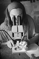
- A stereomicroscope has separate beam paths for both eyes , which show the specimen from different angles, so that a three-dimensional impression is created.
- A line microscope is a reading device on a theodolite , an angle measuring device used in surveying.
- A surgical microscope is used by doctors in the operating room.
- A trichinoscope is used to detect trichinae (roundworms) when examining meat.
- A vibration microscope is used to examine the vibration of strings in stringed instruments.
- A measuring microscope has an additional device that allows the specimen to be measured.
- A computer microscope, for example, can be connected to a computer using a USB cable , which is used to display the image.
history


The principle of magnification using glass bowls filled with water was already described by the Romans ( Seneca ) and magnifying lenses were already known in the 16th century.
Dutch eyewear maker Hans Janssen and his son Zacharias Janssen are often considered to be the inventors of the first compound microscope in 1590. However, this is based on a statement by Zacharias Janssen himself from the mid-17th century. The date is questionable because Zacharias Janssen himself was only born in 1590. In 1609, Galileo Galilei developed the Occhiolino , a compound microscope with a convex and a concave lens. However, Zacharias Janssen had already demonstrated a device with the same functional principle a year earlier at the Frankfurt trade fair . Galileo's microscope was celebrated by the “ Academy of the Lynx ” in Rome, which was founded in 1603 by Federico Cesi . A drawing by academician Francesco Stelluti from 1630 is considered to be the oldest drawing made with the help of a microscope. It shows three views of bees (from above, below and from the side) as well as enlarged details. The bee appeared in the coat of arms of the Barberini family , to which Pope Urban VIII belonged. Stelluti wrote in a banner above the illustration: " For Urban VIII. Pontifex Optimus Maximus [...] from the Academy of the Lynx, and we dedicate this symbol to you with eternal veneration ".
Christiaan Huygens (1629–1695), also Dutch, developed a simple two-lens ocular system in the late 17th century. It was already achromatically corrected, so it had fewer color errors and was therefore a great step forward in improving the optics in the microscope. Huygens eyepieces are still produced today, but they are visually significantly inferior to modern wide-field eyepieces.
Even Robert Hooke used his 1665 published the drawings Micrographia a compound microscope (see figure). The strongest enlargements that he presented in his book were 50 times. Higher magnifications were not possible because the aberrations that occurred in the front lens (objective) and in the eyepiece were multiplied so that no finer details could be seen.

Antoni van Leeuwenhoek (1632–1723) therefore took a different approach. The more curved a lens is, the greater the magnification. Small, approximately spherical lenses therefore have the greatest magnification. Leeuwenhoek was brilliant at precisely grinding the smallest of lenses, a technique that had previously been poorly mastered. His simple microscopes with only one lens were cumbersome to use, but since he only used one lens, there was no need to multiply the aberrations. His microscopes had a magnification of up to 270 times. This is how Leeuwenhoek discovered what he called the “Animalkulen”, unicellular bacteria and protozoa.
In 1768, Michel Ferdinand d'Albert d'Ailly , Duc de Chaulnes (1714–1769) described the first measuring microscope specially designed for measuring purposes .
Robert Brown was still using a simple microscope in 1830 and discovered the cell nucleus and Brownian molecular motion . It took 160 years before compound microscopes produced the same image quality as Leeuwenhoek's simple microscope.
Until well into the 19th century, good compound microscopes were manufactured through trial and error and based on experience. Around 1873, Ernst Abbe worked out the physical principles required to build better microscopes, which are still valid today. As a result, it was possible for the first time to manufacture an objective whose resolution limit was no longer limited by the quality of the material, but by the laws of physical diffraction. This physical limit of resolution is known as the Abbe limit. The corresponding microscopes were produced together with Carl Zeiss in its optical workshops . In doing so, they benefited from the optical glasses developed by Otto Schott and the lighting device developed by August Köhler for Köhler's lighting .
See also
literature
- Jörg Haus: Optical microscopy . Wiley-VCH, Weinheim 2014, ISBN 978-3-527-41127-6 . 220 pages.
- Michael Volger (Ed .: Irene K. Lichtscheidl): Light microscopy online. Retrieved on August 17, 2018 (Theoretical introduction and instructions for practical application at the University of Vienna. Also available as a pdf file (270 pages) .).
- Dieter Gerlach: The light microscope. An introduction to function, handling and special procedures for physicians and biologists . 2nd Edition. Georg Thieme Verlag, Stuttgart 1985, ISBN 3-13-530302-0 .
Web links
How light microscopes work:
- Light microscopy online : Theoretical introduction and detailed instructions for a variety of microscopic techniques on the website of the University of Vienna.
- Various short courses on light microscopy
- Optical Microscopy Primer : Comprehensive tutorial with virtual microscopes
Collections of historical light microscopes:
- Museum of optical instruments: Historical microscopes : Development of scientific microscope construction in Germany with stories about their manufacturers and users, illustrated with over 3000 photos
- Microscope Museum : The history of the light microscope from the beginning until today in words and pictures. Well over 100 microscopes from different manufacturers are presented in the gallery.
Individual evidence
- ↑ a b Gerald Turner: Microscopes . Callwey Verlag, Munich 1981, ISBN 978-3-7667-0561-7 , pp. 25-36 .
- ↑ Dieter Gerlach: History of microscopy . Verlag Harri Deutsch, Frankfurt am Main 2009, ISBN 978-3-8171-1781-9 , pp. 64-110, 171-179 .
- ↑ Hermann Schacht: The microscope and its application . Verlag GWF Müller, Berlin 1851, Chapter: II. 2.
- ^ Zeiss Jena: Catalog No. 30: Microscopes and microscopic auxiliary apparatus . Self-published, Jena 1895, p. 105 .
- ↑ a b c d e Dieter Gerlach: The light microscope. An introduction to function, handling and special procedures for physicians and biologists . Georg Thieme Verlag, Stuttgart 1976, ISBN 3-13-530301-2 , p. 64-71 .
- ↑ a b c d Jörg Haus: Optical microscopy, functionality and contrasting methods . John Wiley & Sons, 2014, ISBN 978-3-527-41286-0 , pp. 17–21 ( limited preview in Google Book search).
- ↑ August Köhler : A new lighting method for microphotographic purposes . In: Journal of Scientific Microscopy . tape X. , No. 4 , 1893, p. 433-440 ( online at archive.org ).
- ↑ Ernst Abbe: Contributions to the theory of the microscope and microscopic perception. In: Archives for microscopic anatomy. 9, 1873, pp. 413-468.
- ↑ German Patent DE 2116521 .
- ↑ Construction plan 1978: Confocal laser scanning fluorescence microscope with high resolution and depth of field / 4Pi Point Hologram (PDF; 83 kB).
- ↑ Computer microscope : The better webcam , test.de, January 23, 2003 (accessed online on February 26, 2013).
- ↑ Stephen Jay Gould : The Lying Stones of Marrakech: Penultimate Explorations of Natural History . S. Fischer, Frankfurt 2003, ISBN 3-10-027813-5 , pp. 52-53 .
- ↑ Hugo Freund, Alexander Berg: History of microscopy. Life and work of great researchers . Volume I: Biology . Umschau Verlag, Frankfurt am Main 1963, p. 4-5 .


