Cell culture
When cell culture is the cultivation of animal or plant cells in a nutrient medium outside the organism referred to. Cell lines are cells of one type of tissue that can reproduce indefinitely in the course of this cell culture. Both immortalized (immortal) cell lines and primary cells are cultivated (primary culture). A primary culture is a non-immortalized cell culture that was obtained directly from a tissue. Cell cultures are widely used in biological and medical research, development and production.
history
Since the beginning of scientific research, there have been efforts to keep cells and tissue alive outside of an organism in order to be able to examine them more closely. Wilhelm Roux succeeded for the first time in 1885 in keeping embryonic chicken cells alive for several days in a saline solution, thus demonstrating the basic principle. In 1913, Alexis Carrel showed that cells can grow longer in cell culture as long as they are fed and kept aseptic .
The oldest animal cell line is believed to be sticker sarcoma , an infectious tumor of natural origin that arose around 200 to 11,000 years ago. Since its inception, sticker sarcoma has accumulated around 1.9 million mutations , and 646 genes have been deleted .
In the years 1951/1952 an immortal human cell line from a cervical carcinoma was established for the first time , which later became known under the name HeLa . In the following decades, nutrient media, growth factors and conditions in particular were further developed and new cell lines were established. In 1975, César Milstein and Georges Köhler discovered the possibility of producing monoclonal antibodies through the fusion of lymphocytes with cancer cells using the hybridoma technique . For this discovery, they received the 1984 Nobel Prize in Physiology or Medicine . In addition, methods for the targeted introduction and expression of genes in cells, the so-called transfection , were developed during these years .
Body cells that have not yet differentiated - so-called stem cells - were first isolated in 1981 from the blastocysts of an embryonic mouse. They tend to differentiate spontaneously in vitro . This can be prevented by factors that promote the self-renewal of the cells. Several such substances have been identified since the late 1980s. Research in this field is currently focused on the cultivation and targeted differentiation of both embryonic and adult stem cells.
principle
Most of the work with cells takes place in a cell culture laboratory . Primary cultures can be created from different tissues , for example from whole embryos or individual organs such as skin, kidneys, etc. The tissue is treated with a protease , for example trypsin , which breaks down the proteins that maintain the cell structure. This causes the cells to become isolated. By adding growth factors , certain cell types can be specifically stimulated to divide. For poorly growing cell types, feeder cells , basement membrane-like matrices and recombinant components of the extracellular matrix are also used.
The tumor cells removed from animal or human tissue are after initial growth on a nutrient medium by analysis of surface antigens ( immunocytology ) or the genome ( PCR and sequencing ) analyzed and selected in order to then bring a large tumor cell clone into culture. The cells can also be genetically modified by introducing a plasmid as a vector . Cells are removed from the stock culture and deep-frozen in liquid nitrogen and are then available for dispatch to other research institutions.
Most cells have a limited lifespan (limited by the Hayflick limit ), with the exception of some tumor-derived cells. After a certain number of doublings, these cells go into senescence and no longer divide. Established or immortal cell lines have acquired the ability to divide indefinitely - either through random mutation (in tumor cells) or through targeted changes (for example through the artificial expression of the telomerase gene).
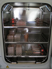
A distinction is also made between cells that grow adherently (on surfaces), such as fibroblasts , endothelial cells or cartilage cells, and suspension cells that grow freely floating in the nutrient medium, such as lymphocytes . The culture conditions differ greatly between the individual cultured cell lines. The different cell types prefer different nutrient media that are specifically composed, for example different pH values or concentrations of amino acids or nutrients. As a rule, mammalian cells grow at 37 ° C with an atmosphere of 5% CO 2 in special incubators. Depending on the rate of division and density of the cells, the cell clusters are loosened every few days and distributed to new vessels (called “passage” or “splitting”). The number of passages indicates the frequency with which the cells have already been passaged. In the case of adherent cells in continuous culture, the cells are regularly isolated in order to avoid confluence and the associated inhibition of cell contact .
Culture media are, for example, RPMI-1640 , Dulbecco's Modified Eagle Medium or Ham's F12 . Balanced salt solutions such as Hanks salts or Earle salts are used for washing and short-term storage (a few minutes) .
application
Cell cultures are widely used, particularly in research and development. The metabolism, division and many other cellular processes can thus be examined in basic research. Furthermore, cultivated cells are used as test systems, for example in the investigation of the effect of substances on the signal transduction and toxicity of the cell. This also drastically reduces the number of animal experiments .
Cell cultures of mammalian cells are also very important for the production of numerous biotechnical products. For example, monoclonal antibodies for research and therapeutic use in medicine are produced using cell culture. Although simple proteins can also be produced in bacteria with less effort, glycosylated proteins must be produced in cell culture, as this is the only place where the correct glycosylation of the proteins takes place. An example of this is erythropoietin (EPO). Many vaccines are also produced in cell culture. Bioreactors are used for the development and implementation of industrial cell culture processes , some in insect cell culture . Disposable bioreactors are of increasing interest for the production of biopharmaceutical products .
In the plant propagation is produced in the Vegetable tissue culture from cell cultures complete plants.
Cell culture lines
It should be noted that the following list of cell culture lines is incomplete. The ATCC alone has up to 4,000 cell lines.
| Cell line | meaning | Origin species | Origin tissue | morphology | link |
|---|---|---|---|---|---|
| 293-T | Contains plasmid with temperature-sensitive mutant of the simian virus 40 large T antigen | human | Kidney (embryo) derivative of HEK-293 | epithelium | DSMZ Cellosaurus |
| A431 | human | skin | epithelium | DSMZ Cellosaurus | |
| A549 | human | Adenocarcinoma of the lungs | epithelium | DSMZ Cellosaurus | |
| BCP-1 | human | blood | lymphocyte | ATCC Cellosaurus | |
| bEnd.3 | brain endothelial | mouse | Brain / cerebral cortex | Endothelium | ATCC Cellosaurus |
| BHK-21 | syrian baby hamster kidney | hamster | Kidney (embryonic) | Fibroblast | DSMZ Cellosaurus |
| BxPC-3 | human | Pancreas , andean carcinoma | epithelium | DSMZ Cellosaurus | |
| BY-2 | Bright Yellow-2 | tobacco | Callus induced on the seedling | DSMZ ( Memento from November 8, 2007 in the Internet Archive ) | |
| CHO | Chinese hamster ovary | hamster | Ovaries | epithelium | ICLC Cellosaurus |
| COS-1 | Originated from CV-1 cells by transformation of an origin-defective SV-40 | Monkey - Chlorocebus aethiops ( Ethiopian green monkey ) | kidney | Fibroblast | DSMZ Cellosaurus |
| COS-7 | Originated from CV-1 cells by transformation of an origin-defective SV-40 | Monkey - Chlorocebus aethiops | kidney | Fibroblast | DSMZ Cellosaurus |
| CV-1 | Monkey - Chlorocebus aethiops | kidney | Fibroblast | Cellosaurus | |
| EPC | herpesviral induced papular epithelioma | Fish ( Pimephales promelas ) | skin | epithelium | ATCC Cellosaurus |
| HaCaT | human adult, calcium, temperature | human | Keratinocyte | epithelium | Cellosaurus |
| HDMEC | human dermal microvascular endothelial cells | human | foreskin | Endothelium | Journal of Investigative Dermatology |
| HEK-293 | human embryonic kidney | human | Kidney (embryonic) | epithelium | DSMZ Cellosaurus |
| HeLa | Henrietta Lacks | human | Cervical cancer ( cervical cancer ) | epithelium | DSMZ Cellosaurus |
| HepG2 | human hepatocellular carcinoma | human | Hepatocellular carcinoma | epithelium | DSMZ Cellosaurus |
| HL-60 | human leukemia | human | Promyeloblasts | Blood cells | DSMZ Cellosaurus |
| HMEC-1 | immortalized human microvascular endothelial cells | human | foreskin | Endothelium | ATCC Cellosaurus |
| HUVEC | human umbilical vein endothelial cells | human | Umbilical vein | Endothelium | ICLC |
| HT-1080 | human | Fibrosarcoma | Connective tissue cells | DSMZ Cellosaurus | |
| Jurkat | human | T-cell leukemia | Blood cells | DSMZ Cellosaurus | |
| K562 | oldest human leukemia cell line | human | blood | myeloid blood cells, established 1975 | DSMZ Cellosaurus |
| LNCaP | human | Prostate adenocarcinoma | epithelium | DSMZ Cellosaurus | |
| MCF-7 | Michigan Cancer Foundation | human | Breast, adenocarcinoma | epithelium | DSMZ Cellosaurus |
| MCF-10A | Michigan Cancer Foundation | human | Mammary gland | epithelium | ATCC Cellosaurus |
| MDCK | Madin Darby canine kidney | dog | kidney | epithelium | ATCC Cellosaurus |
| MTD-1A | mouse | Mammary gland | epithelium | Cellosaurus | |
| MyEnd | myocardial endothelial | mouse | heart | Endothelium | Cellosaurus |
| Neuro-2A (N2A) | Neuroblastoma | mouse | brain | Neuroblast | DSMZ Cellosaurus |
| NIH-3T3 | NIH , 3-day transfer, inoculum 3 × 10 5 cells, contact-inhibited NIH Swiss mouse embryo | mouse | embryo | Fibroblast | DSMZ Cellosaurus |
| NTERA-2 cl.D1 [NT2 / D1] | Pluripotent cell differentiable with tretinoin | human | Testicles , lung metastasis | epithelium | ATCC Cellosaurus |
| P19 | Pluripotent cell differentiable with tretinoin | mouse | Embryonic carcinoma | epithelium | DSMZ Cellosaurus |
| PANC-1 | pancreas 1 | human | Pancreas , adenocarcinoma | epithelium | DSMZ Cellosaurus |
| peer | human | T cell leukemia | DSMZ Cellosaurus | ||
| RTL-W1 | rainbow-trout liver - Waterloo 1 cells | Rainbow trout - Oncorhynchus mykiss | liver | Fibroblast (likely) | Cellosaurus |
| Sf-9 | Spodoptera frugiperda | Insect - Spodoptera frugiperda (moth) | Ovary | DSMZ Cellosaurus | |
| Saos-2 | Osteosarcoma | human | bone | epithelium | DSMZ Cellosaurus |
| T2 | human | T cell leukemia / B cell line hybridoma | DSMZ Cellosaurus | ||
| T84 | human | Colorectal carcinoma / lung metastasis | epithelium | ATCC Cellosaurus | |
| U-937 | human | Burkitt lymphoma | monocytic | DSMZ Cellosaurus |
See also
literature
- Sabine Schmitz: The experimenter: cell culture . 1st edition. Spectrum Academic Publishing House, 2007, ISBN 978-3-8274-1564-6 .
- Toni Lindl, Gerhard Gstraunthaler: Cell and tissue culture. From the basics to the laboratory bench. 6th edition. Spectrum Academic Publishing House, 2008, ISBN 978-3-8274-1776-3 .
- WW Minuth, L. Denk: Advanced Culture Experiments with Adherent Cells. - From single cells to specialized tissues in perfusion culture. Open access publishing. University of Regensburg 2011, ISBN 978-3-88246-330-9 .
Individual evidence
- ↑ C. Murgia, JK Pritchard, SY Kim, A. Fassati, RA Weiss: Clonal origin and evolution of a transmissible cancer. In: Cell (2006), Volume 126 (3), pp. 477-487. PMID 16901782 ; PMC 2593932 (free full text).
- ^ ID O'Neill: Concise review: transmissible animal tumors as models of the cancer stem-cell process. In: Stem Cells (2011), Volume 29 (12), pp. 1909-1914. doi: 10.1002 / stem.751 . PMID 21956952 .
- ↑ a b H. G. Parker, EA Ostrander: Hiding in Plain View-An Ancient Dog in the Modern World. In: Science. 343, 2014, pp. 376–378, doi: 10.1126 / science.1248812 .
- ↑ Landmarks
- ^ ATCC Cell Lines. Retrieved February 6, 2018 .
- ↑ Zbigniew Ruszczak, Michael Detmar u. a .: Effects of rIFN Alpha, Beta, and Gamma on the Morphology, Proliferation, and Cell Surface Antigen Expression of Human Dermal Microvascular Endothelial Cells In Vitro. In: Journal of Investigative Dermatology. 95, 1990, pp. 693-699, doi: 10.1111 / 1523-1747.ep12514496 .
