
Testicles
The testes [ Hodn ] or (more rarely) of / Hode [ hoːdə ] (v. Mittelhochdt . Hode , v. Althochdt . Hodo , v. IE. : Skeu (t) * - "cover, cover") or Testicle (v. Latin : testiculus , Vkl. From testis witness [of virility ], testicle, plural: testes ; - ancient Greek : ὄρχις orchis ), technically also testis and testiculus , is a paired, internal male sexual organ of many sexes reproductive tissue animals . Like the ovaries of female individuals, it belongs to the gonads ( gonads ) and produces the seminal threads ( sperm ). In addition, male sex hormones ( androgens ), especially testosterone , are formed in the testes . In vertebrates, the testes are formed embryonically in the abdominal cavity, but in most mammals they migrate into the scrotum ( scrotum ).

anatomy
Mammals
Size and location

The human testicle is roughly plum-shaped , weighs about 20 grams and has an average volume of 20-25 ml. The average length is 5 cm, the thickness about 3 cm. The testicles do not develop to their full size until puberty and reach their maximum size in the 4th decade of life. With age, the testicle size decreases again. The testicle volume provides information about the functional condition of the testicle, among other things. If the testicle volume is below 8 ml, it can be assumed that the sperm production only works to a limited extent or not at all. Testosterone, on the other hand, is sometimes still produced up to a volume of 1.5 ml; below that, the testicle is usually inoperative.
In mammals , the testicle shape varies from rounded to ovoid. There are clear differences in size, but there is no close relationship to body weight. The largest testicles in the animal world have southern right whales , they make up 2% of the body weight with 500 kg each. Rodents , sheep (up to 300 g each) and domestic pigs (up to 750 g each) have relatively large testicles, while predators are relatively small . In animals with a seasonal period in reproduction, the testicle size is also subject to seasonal fluctuations, the testes are significantly larger in the mating season than in the dormancy period.
In most mammals, both testicles are in sexually mature individuals in the scrotum ( scrotum ) or scrotum-like skin bags. The testicles are created in the abdominal cavity , but migrate through the inguinal canal into the scrotum around the time of birth, in rodents not until puberty . This process is called testicular descent ( Descensus testis ). In some mammals ( e.g. hamsters , bats ) there is a seasonal testicular descent, and the testes are only outside the abdominal cavity during the mating season. However, within mammals there are some groups of animals in which the testes generally remain in the abdominal cavity, the so-called testiconda . The testes can remain at the site of the system (as in elephants ) or they can descend, but still linger in the abdominal cavity (for example in whales , see also table).
| Testiconda | ||
| No testicle descent | Incomplete testicle descent | Seasonal testicular descent |
| Monotremes , golden moles , shrews , Igeltenreks , hyraxes , elephants , sea cows , three-toed sloth , anteaters | Whales , armadillos | Moles , weevils , aardvarks , shrews , bats , some rodents |
Anatomical structure
The external anatomical structure of the testicle is based on the epididymis that adjoins and fuses with it . The testicle section pointing to the head of the epididymis is referred to as the head end ( Extremitas capitata ), the section pointing to the tail of the epididymis as the tail end ( Extremitas caudata ). At the end of the tail there is often a functionless, wart-shaped rudiment of the so-called Müller's duct , which is known as the testicular appendage ( appendix testis , a Morgagni hydatide ). The edge facing the epididymis is the edge of the epididymis ( Margo epididymalis ), opposite it is the free edge ( Margo liber ). In addition, a surface pointing towards the center ( Facies medialis ) and a surface pointing outwards ( Facies lateralis ) can be distinguished.
The testes descend into a bulge in the peritoneum and the inner trunk fascia (referred to here as Fascia spermatica interna ), the processus vaginalis . The process of the vagina is one of the testicular sheaths inside the scrotum. The peritoneum part of this protuberance is called the vaginal skin ( tunica vaginalis testis ). It lines the inside of the scrotum (so-called wall sheet, lamina parietalis or periorchium ), then turns inside as a double lamella and covers the testicles as a visceral lamina ( lamina visceralis or epiorchium ). There is a very narrow space between the two leaves, the cavum vaginale , which ensures that the testicle can move in the scrotum. The connection point between the two leaves is the testicle- cochineal ( mesorchium ), which is used to fix the testicle in the scrotum. The testicle is also at the tail end with a short strip connected to the epididymis (testis own band, ligament testis proprium ). This continues from the epididymal tail as a ligamentum caudae epididymidis and additionally attaches the testicles indirectly to the floor of the scrotum. The testicular lifter muscle ( cremaster muscle ) also attaches to the process of the vagina and acts as a protective device to pull the testicles closer to the abdominal wall when it is touched or cold. In rodents and mammals with seasonal testicular descent, rarely also in individual individuals of other mammals, the muscle can completely retract the testicle into the abdominal cavity (" pendulum testicle ").
A thick whitish connective tissue capsule , the tunica albuginea, lies directly under the peritoneum covering of the testicle . It ensures the mechanical strength of the organ and maintains a certain internal pressure. From this capsule septa pull into the interior and subdivide the testes into testicular lobes ( lobules testis ). The man's testicle has about 350 lobules. The septa also form a body of connective tissue, the mediastinum testis , which in human anatomy is also called the corpus highmori .

Testicles, epididymis and spermatic cord of a male cat :
1 head end
2 tail end
3
edge of the epididymis 4 free edge
5 testicular mesentery
6 epididymis
7 plexus of testicular artery and vein
8 spermatic duct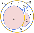
Cross-section through the process of the vaginal membrane:
1 testicle
2 epididymis
3
testicular mesentery 4 organ sheet of the vaginal membrane
( epiorchium )
5 wall sheet of the vaginal skin
( periorchium )
6 cavum vaginale
7 epididymal mesentery
8 internal spermatic fascia
Section through a bull's testicle :
(blood vessels injected with red gelatin )
1 testicular parenchyma
2 mediastinum testis
3 tunica albuginea
4 epididymis tail
5 epididymis head
6 spermatic cord with
tendril convolute of the testicular artery


Vessels and nerves
.jpg)
The testicle is supplied with blood via the testicular artery ( arteria testicularis ). It arises, corresponding to the location of the embryonic attachment of the testicle (see below), directly behind the renal artery directly from the abdominal aorta in the lumbar area. In animals with testicular descent, the testicular artery has to lengthen accordingly and runs along the back of the abdominal wall in a short mesentery ( mesorchium proximal ) to the inguinal canal . Outside the abdominal cavity, it enters the spermatic cord . Here it lies in tight spiral windings, the so-called tendril convolute . In bulls , for example, a two-meter-long artery section is accommodated on a spermatic cord length of 13 cm. The tendril bundle is wrapped in the tendril plexus ( pampiniformis ) of the testicular vein ( vena testicularis ). This creates a large contact area between the incoming and outgoing blood, which acts as a heat exchanger . The temperature in the scrotum is a few degrees below the internal body temperature, which is essential for the formation of fertile sperm in mammals with testicular descent. The incoming blood in the artery is cooled down by this arrangement by the outflowing blood of the vein.
The testicular artery runs along the edge of the epididymis to the end of the tail of the testicle. From there, it moves back to the head end within the testicle capsule at the free edge in most mammals ( ruminants , for example, are an exception ). Their branches are meandering in the tunica albuginea over the side surfaces and pass over the testicular septa inside to the mediastinum testis and from there centrifugally back to the seminiferous tubules, around which they form a capillary network.
The innervation of the testicle is mediated by the sympathetic nervous system , part of the autonomic nervous system . The nerve fibers come from the trunk of the lumbar region and, wrapping around the testicular artery like a braid ( testicular plexus , syn. Superior spermatic nerve ), extend to the testes. A second group of nerve fibers runs from the cloister ganglia of the trunk with the spermatic duct ( ductus deferens ) to the testis ( plexus deferentialis , syn. Nervus spermaticus inferior ). The efferent nerve fibers mainly reach the blood vessels and thus regulate the blood flow and the temperature of the testicle. Participation in the fine control of sperm formation , sperm transport and hormone production in the testes of some mammals is currently being discussed, but this control is primarily carried out via hormones . The cell bodies of those nerve fibers that carry information to the central nervous system ( visceroafferences ) are located in the spinal ganglia of the lumbar region. They transmit pain sensations (intestinal pain ), but a large part of the high sensitivity to pain of the testicle is conveyed via the sensitive nerve fibers of the testicular sheaths (branches of the genitofemoral nerve ). The high sensitivity to touch stimuli makes the testicles and scrotum an erogenous zone . On the other hand, the great sensitivity to pain is also used in BDSM ( cock and ball torture ) and torture ; Squeezing the testicles can lead to shock . A neuralgia of the genitofemoral nerve, which can occur, for example, after surgical correction of an inguinal hernia , manifests itself in testicular pain.
The lymphatic vessels of the testicle run together with the testicular veins to the lumbar lymph nodes ( Lymphonodi lumbales ) backwards of the aorta, in domestic mammals also to the iliac lymph nodes ( Lymphonodi iliaci mediales ) at the aortic branch. In testicular cancer, metastases can occur in these lymph nodes , which are located in the retroperitoneal space of the abdomen .
Remaining chordates
In all other chordates , the testicles are located in the body cavity and below the kidneys.
In the skullless , no compact testicle is formed, the gonads are still segmented, in Asymmetron only the right testicle is present. In hagfish , the elongated testicle is also only one-sided in the form of a lobed band. The round mouths have paired testicles that extend the entire length of the body cavity.
Within the cartilaginous fish , only in the collar shark , the anatomically and morphologically most primeval shark, does the entire testicular system develop into an elongated gonad, in the other representatives, as in the other vertebrates , only the middle section (see below). The testicles of the real sharks are also elongated, those of the rays are short and flat. In dogfish and electric rays , the testicles are far in front, near the pericardium , otherwise in the middle or rear part of the trunk. In most bony fish , the testicles are elongated. In the real bony fish they are located below the kidneys and the swim bladder and are attached by a mesorchium. In some perch species , both testicles are fused together at the rear end. The thin tunica albuginea is occasionally pigmented in bony fish . At the same time, ovarian tissue can appear in the testicles of fish (hermaphrodite, ovotestis ). Sawfish and sea bream are hermaphrodites , so they have both testicles and ovaries and can change sex in the course of their lives.
Amphibians have either elongated ( tail amphibians , crawling amphibians ) or roundish ( frogs ) testicles. They are attached to the trunk wall or the urnal kidney via a mesorchium . A distinct fat body is formed medially (in the direction of the median plane ) of the testicle. In salamanders , several testicular departments are fused into one flap, the number of departments increases with age. In amphibians, the urinary and seminal tract is beginning to separate. The epididymal ducts ( Ductuli epididymidis ) open into the Wolff duct just before the cloaca . Male toads are hermaphrodites. In front of the testes lies the bidder organ , a primitive ovary, which develops from the anterior gonadal anlage .
In reptiles , the testes are in front of and below the kidneys in close proximity to the adrenal glands . The testicles are oval, more rounded in tortoises and elongated in snakes . The epididymis are medial (towards the center of the body) of the respective testicle. The connective tissue framework of the testicle is poorly developed.
In birds , too , the testes lie in front of the kidneys on the adrenal glands. In contrast to the ovary, which is only one-sided in birds, the testes are always paired. The testicle size shows the strongest seasonal fluctuations within the vertebrates, it increases, for example, in passerine birds by up to 800 times during the mating season and decreases again during mating rest. In a house drakes the testes are in the mating season about 8 cm long and 4.5 cm wide. The connective tissue framework of the testicle is only slightly developed in birds, the tunica albuginea is thin, and there is no mediastinum testis . The endoscopic examination of the testicle used to play a major role in determining the sex of species that do not show any sexual dimorphism , but today it has largely been replaced by molecular biological methods.
Invertebrates
There is asexual and sexual reproduction within the cnidarians . During sexual reproduction, the germ cells are released into the surrounding water or gastric space by bursting the epithelium . Rib jellyfish are generally hermaphrodites and have testicles and ovaries embedded in the mesogloea under the comb-like plates ("ribs") .
Primordial mouths (protostomia)
Most flatworms (platelets) are hermaphrodites. They do not have a body cavity ( coelom ), the testes lie adjacent to the ovaries in a connective tissue space ( interstitium ) within the animal, in tapeworms within each tapeworm limb ( proglottid ). Some flatworms ( Catenulida and Acoelomorpha ) do not have gonads. Also belly-haired (Gastrotricha) do not have a coelom, the freshwater species reproduce asexually via parthenogenesis . The salt water representatives are hermaphrodites, but only the gonads of one sex are functionally active. The germ cells are released through piles of sperm . Rotifers (Rotatoria) have a pseudocoeloma with paired or unpaired testicles, in some species so-called dwarf males occur, in others no males, the reproduction then takes place via virgin generation. Cycliophora reproduce asexually in the so-called feeding stage. In sexual reproduction dwarf males occur. They have two external "testes" and a copulatory organ next to the adhesive disc. In scratch worms (Acanthocephala) a so-called pulling genital ribbon through the body, at the lower third of the two balls seated. A sperm duct runs from each testicle to the penis.
Priapulida and corset animals (Loricifera) have paired testes that are united with the protonephridia to form a urinary and sexual apparatus . With roundworms (Nematoda) there are both separate-sex species and hermaphrodites. The elongated testicle lies below the intestine and also opens into it (see Fig.). String worms (Nematomorpha) have paired testes in the pseudocoeloma. Tardigrade (Tardigrada) are separate sexes. While the body cavity represents a pseudocoelom, there is a real coelom around the unpaired testicles. Colibus (Onychophora) have paired testes that open into a common spermatic duct via ducts. In the arthropods (Arthropoda: millipedes , insects , crustaceans and chelicerates ) males have paired testes in the blood-filled pseudocoel ( haemocoel ) of the abdomen or the rear trunk segments. In some arthropods there are also hermaphrodites.
The annelids have paired testicles. Belt worms are mostly hermaphrodites, but fertilize each other. The testes are located in the coelom , in earthworms in the 10th and 11th body segment and the spermatic duct opens outward in the 15th segment. Multi-bristles , on the other hand, are mostly unisexual and male representatives have testicles in every body segment. In molluscs (Mollusca) there are hermaphrodites, hermaphrodite gonads ( Ovotestis ) and separate sex forms. The coelom is reduced to two cavities around the gonads ( gonadocoel ) and around the heart behind ( pericardium ). In armpods (Brachiopoda) the testes lie in the coelom (more precisely in the metacoel ), the gametes are derived via the metanephridia .
Neumünder (Deuterostomia)
Within the echinoderms (Echinodermata), sea whales have only one testicle, in sea urchins and sea whales the testicles fill almost the entire metacoel, in sea lilies (three to five gonads) and starfish (two per arm) they lie in the arms and open between them Arms with one genital opening each.
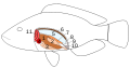
Internal organs of a bony fish:
7 testes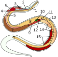
Internal organs of a snake:
14 testes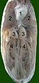
Site of a domestic cock:
(excluding digestive organs)
3 testes
Roundworm:
5 testes
Blueprint of a pod with gonads in the metacoel


Fine structure and function in vertebrates
The testicular lobes each contain two to four coiled seminiferous tubules ( tubuli seminiferi contorti s. Convoluti ), which represent the testicular parenchyma . They are about 50 to 80 cm long and 150 to 300 µm in diameter. Its wall consists of a connective tissue shell with contractile myofibroblasts , a basement membrane and the germinal epithelium ( epithelium spermatogenicum ).
This epithelium consists of sperm or germ cells ( Cellulae spermatogenicae ) and Sertoli cells . The sperm cells are formed from the germ cells ( spermatogenesis ). Since spermatogenesis is the most important task of the testicle, the germ cells are also most frequently present in the testes in terms of quantity. During sperm formation, the successive developmental stages of the germ cells ( spermatogonia → spermatocytes → spermatids → sperm) are gradually transported towards the lumen . Sperm formation takes between 35 (mouse, pig) and 64 days (human), but then further maturation in the epididymis is necessary so that the sperm can be fertilized. This lasts a week in most mammals and 8 to 17 days in humans. In men, around 200 to 300 million sperm are released from the epididymis per ejaculation . With more frequent ejaculation, the amount of sperm will decrease because the daily sperm production capacity is limited. It depends on the testicle mass and the number of Sertoli cells and is between 45 and 200 million sperm per day in men.
The second important component of the seminiferous tubules are the Sertoli cells ( Epitheliocyti sustentantes ). They are about 70 to 80 µm long and run radially through the entire germinal epithelium up to the lumen. The Sertoli cells have a supporting and nursing function for the sperm cells, they nourish the sperm cells, ensure their correct hormonal environment and use plasma movements to transport them to the lumen. In addition, the Sertoli cells phagocytose degenerated sperm cells and cell debris that arise during sperm development. The Sertoli cells are activated by the follicle stimulating hormone of the (FSH) pituitary controlled, its distribution on the formation of the hormone inhibin B influence. They also secrete the androgen binding protein , the anti- Müllerian hormone and a potassium- rich seminal fluid.
The Sertoli cells have numerous processes that surround the germ cells. These cell processes connect at the base of the seminiferous tubules via tight junctions with those of neighboring Sertoli cells and thus form what is known as the blood-testis barrier . This term is actually misleading because this barrier does not lie between blood and testicular tissue, but runs between the spermatogonia and spermatocytes, thus dividing the testicular tubules in a circular manner into a basal and a lumen-facing (adluminal) compartment . The blood-testicle barrier is impermeable to most proteins and protects the sperm from mutagens and the body's own defenses . The latter is necessary because the first sperm cells only emerge after the lymphocytes have been imprinted (see self-tolerance ), so the immune system would consider them to be foreign cells. However, anti-inflammatory cytokines , the release of which is presumably androgen- dependent, and the cells of the immune system in the testes ( dendritic cells , macrophages ) also play a role in protecting against autoimmune reactions .
In many vertebrates, the convoluted seminiferous tubules merge into a short straight seminiferous tubule ( tubulus seminifer rectus ) at both ends . The straight tubules are lined by a single-layer epithelium and open into a canal system in the mediastinum, the testicular network ( rete testis ). The canal system of the testicular network is also mostly lined by a single-layer epithelium (two-layer in cattle). In men, stallions and rodents, however , the testicular network is mainly on the periphery of the testicles ("extratesticular rete"). Several meandering ductuli efferentes testis run from the testicular network into the epididymal head and unite there to form the epididymal duct. In mammals there are about 15 efferent ductules, the number varies within vertebrates between one (e.g. rays ) and 32 (e.g. axolotl ).
The tissue between the convoluted seminiferous tubules is called the interstitium . In most vertebrates it makes up about 10 to 20% of the testicular tissue, in extreme cases such as the woodchuck almost 70%. In addition to connective tissue, blood vessels and nerve fibers , the interstitium also contains Leydig cells ( Endocrinocyti interstitiales ). Via special cell contacts ( gap junctions ) they form interconnected cell associations, so-called functional syncytia . The Leydig cells produce, depending on the luteinizing hormone (LH), male sex hormones (androgens such as testosterone and androstanolone ) and oxytocin , which promotes the motility of the seminiferous tubules. The testicle is thus also an endocrine organ . Testosterone causes the spermatids to mature in the seminiferous tubules. In order to get through the blood-testicular barrier to its place of action, it needs the androgen-binding protein of the Sertoli cells. The androgens also have a variety of effects in the body, including promoting the development of secondary sexual characteristics , having an anabolic effect and controlling sexual behavior . In addition, the Leydig cells form numerous other hormonally active peptides that act on neighboring cells ( paracrine ) or on the Leydig cell itself ( autocrine ) that make them up.

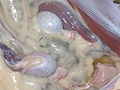
Histological picture of the testicular parenchyma of a boar:
1 lumen of a convoluted seminiferous tubule
2 spermatids
3 spermatocytes
4 spermatogonia
5 Sertoli cells
6 myofibroblasts
7 Leydig cells
8 capillaries
Germ epithelium of the seminiferous tubule :
1 basement membrane
2 spermatogony
3 spermatocyte 1st order
4 spermatocyte 2nd order
5 spermatids
6 mature spermatids
7 Sertoli cells
8 tight junction
(blood-testicle barrier)
Scheme of the internal structure of the testicle and the epididymis :
1 tunica albuginea
2 septa
3 testicular lobes
4 mediastinum testis
5 convoluted seminiferous tubules
6 straight seminiferous tubules
7 rete testis
8 ductuli efferentes testis
9 epididymis
10 beginning of the spermatic duct


Hormonal control

The hormonal control of the testis is carried out by fits and starts, by nerve cells in the median eminence in the hypothalamus formed gonadoliberin (GnRH). GnRH does not act directly on the testes, but stimulates the formation of the hormones LH and FSH in the anterior pituitary gland . The release of these hormones is also controlled by the testes themselves via a negative feedback mechanism: FSH secretion is inhibited by the inhibin B produced by the Sertoli cells, while GnRH secretion is inhibited by the testosterone produced by the Leydig cells . The seasonal fluctuations in the size and activity of the testes in many animals are mediated by the suppression of GnRH secretion during rest under the influence of the length of daylight. The exact mechanism is not yet known in detail: In mammals, opioids , dopaminergic neurons and melatonin are presumably involved in this control loop, in birds also the thyroid hormones .
LH binds to a membrane receptor in the Leydig cells and thus induces the synthesis of androgens . In this case, cholesterol gradually, among other things, pregnenolone and progesterone , converted to testosterone, using two different synthetic routes (Δ4 and Δ5) are possible. The LH-effect on the Leydig cells is prolactin potentiated, but with an overproduction of prolactin occurs by down-regulation of LH receptors to inhibition of testosterone synthesis. LH can also induce the formation of androgens in the adrenal cortex ; the dehydroepiandrosterone formed there reaches the testes via the blood and can be used there as a testosterone precursor. About 97% of androgens are formed in the testes (about 7 mg / day in men), the remaining part in the adrenal glands . Androgens act on the germinal epithelium and are bound to a protein and transported via the blood to their other target organs.
FSH binds to the corresponding receptors on the Sertoli cells. Both FSH and testosterone control spermogenesis. FSH initiates spermiogenesis, testosterone promotes mitotic and meiotic cell division and thus the formation of spermatocytes from the spermatogonia, while FSH in turn causes the final maturation of the spermatids to sperm.
The suppression of the hormonal stimulation of the testicular function is currently being intensively researched in the development of contraceptives for men . Testosterone or its combination with GnRH antagonists or gestagens such as progestin are currently viewed as the most promising candidates. They lead to greatly reduced testosterone concentrations within the testicle and thus to a strong or complete reduction in sperm formation. In veterinary medicine, a preparation based on the GnRH analogue Deslorelin ( Suprelorin ® ) has been approved since 2008 , which suppresses fertility in males for several months. In addition, a vaccine for pigs ( Improvac ® ) is approved, which leads to the formation of antibodies against GnRH and thus suppresses testicular function.
Development history

The prerequisite for sexual reproduction is the separation of the cells specialized for reproduction (germ cells) from the normal body cells (somatic cells). This separation is already carried out in ciliate animals in the form of a micronucleus , more clearly then in spherical algae , where the main cell group of body cells is opposed to a small group of germ cells ( gonidia ), which, however, is not yet developed in the form of a delimited organ . The presence of testes (or, in principle, of gonads) is not a basic characteristic of multicellular cells.
In the Bilateria , a third cotyledon , the mesoderm , and thus complex organs , appear for the first time . However, in many invertebrates sexual reproduction is still combined with the possibility of asexual reproduction. This often involves a generation change , i.e. the sexual one follows an asexual reproductive cycle.
The differentiation of the gonads into testes and ovaries is a hallmark of segregated species. So far it has not been clarified whether hermaphroditism or segregated sex is the plesiomorphic characteristic of bilateria. Both gonads emerge from the same system in embryonic development. In many animal groups, despite this gender separation, reproduction without fertilization ( parthenogenesis ) is possible, which can be viewed as a reduced form of sexual reproduction. Male animals only appear here in exceptional cases. Parthenogenesis is found in numerous taxa , from rotifers to some lizards . Up to the amphibians, hermaphroditic forms or a change in gender ( dichogamy ) can also be found during ontogenesis . It is possible to convert the ovaries into testes ( proterogyny ) and the testes into ovaries ( proterandry ).
In most animals, it is genetically determined whether the sexually indifferent structure of the gonads results in a testicle or an ovary . In worms and flies, the sex is determined by the ratio of X chromosomes and autosomes . In state-forming insects, testes develop from unfertilized eggs in the offspring, and ovaries in animals with a diploid set of chromosomes, i.e. from fertilized eggs. In mammals, sex is determined by the Y chromosome . A gene ( sex determining region of Y , Sry ) is located on this sex chromosome ( gonosome ) , which interacts with genes of other chromosomes and (in humans from the 7th week after fertilization) leads to the formation of the testicular-determining factor . This initiates the development of the testicles and thus the male sex in general. It encodes a number of transcription factors , the so-called HMG proteins ( high mobility group proteins ). These proteins have numerous other functions in other tissues, and the precise mechanisms involved in testicular formation are currently being intensively researched. With the expression of Sry, the Sertoli cells differentiate, which among other things produce the anti-Müllerian hormone and thus cause the regression of the Müllerian ducts . The further development of the testicle and that of the other characteristics of the male sex is controlled by androgens. For some animal groups, however, the sex is determined by environmental factors. In some amphibians and many reptiles ( turtles , alligators ) , for example, the sex depends on the incubation temperature. ( See also. Temperature-dependent sex determination )
In many animals, the gonads are developed in close relationship with the excretory system ( primitive kidney , nephridia ), especially the urinary tract be shared as samenableitendes system, which is why both organ systems in vertebrates as urinary and sexual apparatus summarizing. In invertebrates, however, the place of origin, location and ducts are very different, so that it is assumed that sexual reproduction has arisen multiple times and independently of one another in evolution . The complexity of the sexual organs does not depend on the stage of evolution; it is very high in flatworms, for example.
Vertebrate embryology
Testicular development in vertebrates
|

In the embryo , the testes and ovaries arise from the same system , the so-called genital ridge . It forms in the area of the urnal kidney and initially extends from the thorax to the loin . In most vertebrates, only the middle part of this elongated system becomes the actual gonad, the remaining sections develop into the gonad ligaments . Under the influence of Sry, the primordial germ cells from the yolk sac migrate into the gonadal system (in humans in the 6th embryonic week) and the epithelium of the primitive body cavity ( coelom ) grows finger-like into the system as so-called primary germ lines .
The germ or testicular cords penetrate the gonadal system and grow around the primordial germ cells. This temporarily divides the gonads into cortex and marrow, although in male embryos only the marrow develops into the testicle, while the cortex is regressed. Similar processes take place in genetically female individuals, but later and the ovary is formed from the cortex while the marrow degenerates. The connection between the testicular cords and the surface is eventually lost. From the testicular cords, the Sry-expressing pre-Sertoli cells develop into the Sertoli cells , which are regarded as the organizers of further testicular development and thereby interact with the myoid cells . The spermatogonia arise from the primary sex cells .
Inside, the testicular cords form a network of interconnected cords, the later testicular network ( rete testis ). The testicular network connects to some urnal tubules, which thus become the efferent ducts of the head of the epididymis . The urinary duct ( Wolff duct ) is also converted into a sperm drainage path as an epididymal canal and spermatic duct . However, the lumen of the seminiferous tubules does not develop until puberty , in amphibians after metamorphosis , until then the testicular cords are solid.
The tunica albuginea , the connective tissue framework of the testicle and the Leydig cells arise from the mesodermal portion of the testicular anus . The Leydig cells can also be found in the early testicular development; they express steroidogenic factor 1 (Sf1), and their testosterone production largely determines the development of the male sexual organs.
Aging
In men - in contrast to women ( see menopause ) - there is no sudden cessation of the function of the gonads at a defined age. Both the hormone production and the maturation of the germ cells are potentially preserved into old age. In fact, paternity has been documented up to the tenth decade of life . In reality, however, there are marked individual differences and many men sooner or later become infertile , which should not be confused with impotence . The exact causes for these differences are not known in detail; vascular factors are suspected, among others . Statistically, it comes from about the fourth decade of life to the very slow pace of involution of the testes with loss of weight, size and sperm production. For the individual, however, there are hardly any predictions to be made.
The structural changes are therefore also subject to a wide range, but a mixed picture of normal and clearly atrophic testicular tubules can be regarded as typical . A significant decrease in testosterone production observed in some men can lead to Climacterium virile with hot flashes , headache and other symptoms .
History of exploration

In antiquity and the Middle Ages, the testicle was only considered a transit station for the semen. Alkmaion von Kroton (early 5th century BC) suspected the brain as the origin of the semen, which reached the testes via blood vessels . The atomists ( Anaxagoras , Democritus ) and Aristotle included the spinal cord in this path, Galenus (125–199) suspected the origin of the sperm cells in the blood vessels through which they reach the testes. These ideas persisted into the Middle Ages. Leonardo da Vinci's anatomical drawings show connections between the testicles and the lungs and brain, because da Vinci suspected that the “spiritual power” of the semen came from the brain, while the testicles only provide the material basis for the “lower impulses”.
Structural exploration
It was only with the beginning of the Enlightenment in the 17th century that the ideas of male semen were demystified and the direct connection between testes and reproduction recognized. The first modern description of the structure of the testicle comes from Reinier de Graaf (1641–1673). Nathaniel Highmore described in 1651 the connective tissue body of the testicle ( Corpus Highmori ), which was called Mediastinum testis by Astley Paston Cooper in 1830 . In 1677, the inventor of the microscope , Antoni van Leeuwenhoek , discovered sperm, which he believed to be miniaturized pre-formed organisms ("seed animals").

With the development of histological techniques, the fine structure of the testicle could also be clarified. In 1841, the Swiss anatomist Albert von Koelliker recognized the direct connection between testicular tubules and sperm for the first time and discovered that the sperm arise in these tubules as products of cellular differentiation. In 1850, Kölliker's pupil Franz von Leydig first described the intermediate cells ( Leydig cells ).
In 1865 Enrico Sertoli discovered the supporting cells ( Sertoli cells ). In 1871 Victor Ebner succeeded in differentiating the Sertoli cells from the spermatogonia and five years later La Valette St. George coined the term "spermatogony" and the division of the individual developmental stages of the sperm cells, which is still common today. The Sertoli cells were long regarded as syncitia , it was not until 1956 that Don W. Fawcett and Mario H. Burgos were able to prove that each Sertoli cell has its own cell boundaries.
Hugo Ribbert recognized as early as 1904 that carmine administered into the blood did not get into the lumen of the seminiferous tubules and the testicular network. For a long time, this discovery was neglected, although it was the first evidence of the blood-testicular barrier. This knowledge was only taken up in the late 1950s and in 1963 J. Brökelmann succeeded in proving the tight junctions of the Sertoli cells as the morphological basis of the blood-testicular barrier. A year later, Paul J. Gardner and Edward A. Holyoke were able to explain the fine structure of the blood-testicle barrier.
Hormones
Although the effects of castration had been known for thousands of years, it was not until 1849 that Arnold Adolph Berthold succeeded in experimentally demonstrating the formation of hormones in the testes by means of testicular transplants in cocks. At a very old age, Charles-Édouard Brown-Séquard undertook self -experiments at the end of the 19th century with fluid from the testes of dogs and guinea pigs, which he ascribed a rejuvenating and strengthening power, although it was more homeopathic hormone quantities that he obtained in this way. At the beginning of the 20th century, the transplantation of animal testicles under the abdominal wall was considered a means of rejuvenation, in particular the Viennese Robert Lichtenstern and Eugen Steinach were protagonists of this method. Steinach wanted to achieve this rejuvenation process by ligating the vas deferens (his most famous patient was Sigmund Freud ) and described testicular transplantation as a “therapy” for homosexuality . After 1945, these controversial xenografts went out of fashion.
In 1903, Pol Bouin and Paul Ancel first published the knowledge that the Leydig cells are the place where male sex hormones are produced. In 1931 Adolf Butenandt and Kurt Tscherning isolated androsterone (a metabolite of testosterone) from the urine of men, in 1935 Ernst Laqueur was able to isolate testosterone himself from bull testicles and also coined the name of this hormone (from testis "testes" and " steroid ").
The existence of non-steroidal hormones in the testes was postulated as early as the 1920s, but it was not until 1932 that D. Roy McCullagh functionally demonstrated it in castrated rats and called it inhibin . Although bioassays for this hormone were developed in the 1960s , its existence was controversial for several decades and was not widely accepted until 1979. In 1984/85 the structure and subtypes of the hormone were revealed. With the clarification of the DNA sequence coding for the hormone , the affiliation of the inhibin to the group of β-transforming growth factors was recognized in 1985 .
While the relationships between LH and testosterone were already known in the 1960s, the FSH dependence of the Sertoli cells was only proven in 1984 by Joanne M. Orth .
Gender differentiation
The chromosomal basis of sex differentiation was already cleared up between 1910 and 1916, primarily through the work of Thomas Hunt Morgan on fruit flies , for which he received the 1933 Nobel Prize in Physiology or Medicine . Alfred Jost recognized in 1947 that the gonads are primarily determined by the female gender and that the male gender is dependent on testosterone. Nevertheless, it was not until the early 1960s that the Y chromosome was identified as a major factor in mammals. The exact localization of the gene for the testicular-determining factor was not determined until 1990, its various functions are not yet known in detail and are the subject of current research.
Developmental disorders and diseases
Injuries to the testicle occur in humans as blunt trauma , especially in martial arts and fights. There is a risk of bleeding under the testicle capsule ( hematocele ), which usually requires surgical treatment. Injuries with opening of the scrotum (puncture and impaling wounds, in animals also bites, barbed wire, etc.) can cause testicular inflammation (see below) or even abscesses , as well as peritonitis due to the open connection of the vaginal process to the abdominal cavity .
Malformations
An anorchy is the absence of both testicles; if only one testicle is formed, one speaks of a monorchy . About 5% of male children operated on for a lack of testicular descent have only one or no testicle. They often have small connective tissue nodes with scattered Leydig cells. Since a functionally intact testicle is imperative for the male gender expression, at least one intact testicle must have been present in the embryonic phase, which may have regressed later.
In rare cases, as a result of developmental disorders, ovaries can also appear in addition to testicles in humans and other mammals ( hermaphroditism verus - "real" hermaphrodites; see also intersexuality ). With certain malformations of the gonads ( gonadal dysgenesis ), the testicles are not created, remain underdeveloped or contain ovarian tissue ( ovotestis ).
A very rare undesirable development is the splenogonadal fusion with a connection between testicular and spleen tissue .
With disturbances of the migration of the testicle ( Maldescensus testis ) various positional anomalies can occur. The testicle can remain in the abdominal cavity ( cryptorchidism , "abdominal testicle "), get stuck in the inguinal canal ("inguinal testicle", "glide bone") or take a wrong route and, for example, come to lie under the skin of the groin or the inner thigh ( testicular ectopia ) . The Maldescensus testis is one of the most common malformations in humans and occurs in 3 to 5% of newborns and 33% of premature babies; cryptorchids also occur with a similar frequency in domestic animals and lead to exclusion from breeding. If the testicle is incorrectly positioned, fertile sperm cannot form due to the temperature sensitivity of the germinal epithelium, but androgen production is retained. Failure to descend for more than two years can lead to the loss of spermatogonia and thus irreversible changes in the testicle.
Hereditary short stature of the testicles ( testicular hypoplasia ) is relatively common in pets. Testicular hypoplasia can also be caused by chromosomal disorders ( Klinefelter syndrome ), infections, or hormonal disorders .
An abnormal enlargement of the testicles is called macroorchidy .
Inflammation of the testicles
Inflammation of the testicles ( orchitis ) can occur with injuries to the scrotum with invasion of bacteria or with some infectious diseases. Orchitis is a possible complication of mumps , Coxsackie virus infections, and chickenpox in humans . Also brucellosis and tuberculosis may manifest on testicles. In animals also tuberculosis and can brucellosis and pseudotuberculosis ( sheep ), the equine infectious anemia and feline infectious anemia ( cats associated) with a Orchiditis. Inflammation of the testicles can lead to shrinking of the testicles ( testicular atrophy ) and infertility , because no more sperm cells ( aspermia ) or no functioning sperm can be formed.
Circulatory disorders
As a varicocele is called varicose veins similar extensions that especially the left side veins of the plexus pampiniformis concern in the spermatic cord. A varicocele can lead to limited sperm production in the testicle on the same side.
The accumulation of serous fluid in the testicular sheaths is called a hydrocele or water break. The fluid can also collect in the spermatic cord, which is then referred to as the hydrocele funiculi spermatici .
A torsion is an abnormal rotation of the testis, wherein said helical-clamping of the spermatic cord and the draining veins can lead to the death of the testis. Severe testicular torsion is a very painful emergency, and permanent damage to the testicle can be expected after just two hours. The testis appendix can also develop a so-called hydrate torsion.
Circulatory disorders with the risk of developing necrosis are also observed in diseases of the blood vessels such as Henoch-Schönlein purpura , endangiitis obliterans and panarteritis nodosa in humans, arteritis in horses and generally in thrombosis .
Tumors
A testicular tumor is a pathological enlargement of the testicle. Testicular tumors can be benign or malignant .
Mostly harmless testicular enlargements are cysts . Two different types of cysts can develop on the testicle. Hydroceles are sacs of the tunica vaginalis testis that contain a clear, amber-colored liquid. They are caused by injury or inflammation. Spermatoceles originate from the rete testis or the epididymis and contain sperm. Teratomas are mostly benign tumors of the germ cells.
A very rare cause of a mass on the testicle can be splenogonadal fusion .
Malignant testicular tumors ( testicular cancer ) are subdivided into degenerations of the germ cells ( germinal testicular tumors : seminomas ) and non- seminomas . Degeneration of the germ cells is the most common cancer in men between the ages of 20 and 40 and accounts for around 90% of all testicular tumors. The main risk factor are testes that have not migrated into the scrotum. The remaining 10% are due to tumorous degeneration of other tissue parts ( Sertoli cell tumor , Leydig cell tumor , non-Hodgkin lymphoma, etc.).
Malfunctions
In addition to the diseases mentioned above , chemical substances that overcome the blood-testicular barrier such as environmental toxins (e.g. cadmium ), additives to packaging (e.g. phthalates , diethylhexyl adipate ), some drugs (e.g. furazolidone ) and hormones can also occur (see also endocrine disruptors ) or ionizing radiation lead to severe impairment of the epithelium of the seminiferous tubules. Since sperm formation is associated with very high rates of cell division ( mitosis , meiosis ), the germinal epithelium is particularly sensitive to cell toxins. Such damage can lead to manifold changes up to the complete absence of sperm ( see also sperm analysis ).
Inadequate production of androgens is known as hypogonadism . This can be congenital, secondary to diseases of the testicle or a gonadotropin deficiency (e.g. hypofunction of the pituitary gland , olfactogenital syndrome ).
Abdominal cryptorchidism in a domestic sheep ("kidney buck")

Testicular hypoplasia in a cat, on the right the normally developed testicle.

Distinct varicocele on the left.

Dog testicles removed after rotation (left: healthy organ)

Sonography : dog with abdominal cryptorchidism and ascites (left: testicles, right: epididymis)


examination
The testicular examination is an important basic examination for humans and animals with a scrotum. Here the presence, size, location and consistency of the testicle are checked. The ultrasound examination is mainly used as an imaging method . The testicular volume is determined either by comparison with the so-called Prader chain or by means of ultrasound measurement. Diaphanoscopy still has a certain value in the diagnosis of hydrocele . A testicular biopsy can be performed to take tissue samples . In animals with testicles in the abdominal cavity, endoscopy is used in addition to the ultrasound examination .
A functional examination is the creation of a semen analysis . Here the number, shape and mobility of the sperm are assessed. The determination of the level of inhibin B in the blood is used as a marker for Sertoli cell function and fertility, but its informative value is controversial.
The Leydig cell stimulation test can be used to detect testicular tissue that cannot be detected using imaging techniques .
castration

The restriction of testicular function is called castration. It can be done by surgical removal of the testicle ( orchidectomy ), ligation of the testicular vessels ("bloodless castration"), radiation or chemical substances. Castrations are mainly carried out in humans for testicular cancer. Surgically removed testicles are usually replaced by a testicular prosthesis for cosmetic reasons .
As a symbol of disempowerment, castration also plays a role in the mythology of many cultures (see also castration fear ). In Egyptian mythology, Horus tears the testicles from his half-brother Seth . In Greek mythology , Kronos first removes the testicles of his father Uranus and is later emasculated by his son Zeus himself . To renounce worldly desires, the self- emasculation of the Galloi (priests) was common in the Cybele cult of the Phrygians , which also spread to ancient Greece and Rome, as was the case with the Hijras in India . In Judaism, however , castration, both of humans and animals, is strictly prohibited. In Christianity , castration was also frowned upon. Eunuchs were not allowed to be ordained priests, but there were currents in which self-castration was performed as a ritual (see Skopzen ).
Historically, slaves , prisoners of war, singers or the guards of harems (see Palasteunuch ) were also castrated. The non-medically justified castration was aimed in particular at the prevention of the secondary sexual characteristics (pitch of the voice, sexual behavior) caused by the testosterone. Castrati were popular in European musical life in the 17th and 18th centuries and were often highly regarded. Senesino , Farinelli , Caffarelli and Antonio Bernacchi are among the most famous castrati of the 18th century .
Castrated men cannot reproduce by themselves. Similar to clerics who voluntarily abstained ( eunuchs for the kingdom of heaven ), they were judged to be more reliable and used in various societies as functionaries and servants. The voluntary castration of sex offenders is still a therapy method, albeit a controversial one, in Germany and in some states of the USA.
In veterinary medicine, castration is used, in addition to medical indications (testicular cancer, prostate and anal gland diseases), above all to avoid offspring, to improve the manageability of pets ( gelding , ox ), to increase fattening performance and meat quality, in domestic pigs also to avoid "Boar smell" of the meat carried out. Castrations on animals were probably carried out as early as the beginning of the Neolithic . Castration is one of the few non-medically indicated organ removals still permitted in Germany today under the Animal Welfare Act (§ 6), in very young animals even without eliminating pain, although this is not undisputed.
Cultural and historical significance

In Japanese mythology , Tanuki , demons ( yōkai ) similar to the raccoon dog , are often represented as a symbol of good luck with oversized testicles. In ancient Greece , the genitals of animals, especially bulls ( taurobolium ), were offered as sacrifices. Testicles were considered a symbol of virility and creative potency. According to Taylor, they had a stronger symbolic power than the penis until the late 16th century .
botany

The plants, which in their appearance resemble male genitalia, were thought to have an aphrodisiac and fertility-increasing effect. The Greek philosopher Theophrastus von Eresos named them Orchis , the Greek name for testicles, due to the similarity of the two bulbs of the orchid with the testicles. Their consumption is said to be conducive to the birth of a boy ("orchid"). Orchis later gave its name to the entire orchid family .
The name avocado is derived from the Indian word ahuacatl (testicle), which refers to the testicle-like shape of the fruit of this tree.
In art
In art, in contrast to the phallus , testicles do not play a central role outside of erotic and pornography . “Blood and Testicles”, a phonological play on words on the blood-and-soil ideology , is often used derogatory in art criticism .
One of the characters in Thomas Mann's Tristan is Mr. Klöterjahn (“Klöten” is the Low German expression for testicles, a symbol of fitness for life and vitality). In the novel Sanningen om Sascha Knisch (German title The Truth About Sascha Knisch. 2003) by the Swedish author Aris Fioretos , the testicles are the leitmotif . The German film Eierdiebe deals with the subject of testicular cancer and the loss of a testicle.

The coat of arms of the Italian noble house Colleoni from Bergamo shows several pairs of testicles and is probably an allusion to coglione , an Italian name for testicles. A statue of Bartolomeo Colleoni with this coat of arms on the base prays the main character in the first volume of Heinrich Mann's three-part novel The Goddesses or The three novels of the Duchess of Assy .
As food

Testicles are processed as food in many regions. Germany was the only country in the EU where testicles were banned as food. According to EU regulation no. 853/2004 of April 29, 2004, testicles are the only genital organs considered food, all others are classified as unsuitable for consumption ( confiscates ).
Others
- The American Gregg Miller received the Ig Nobel Prize in Medicine in 2005 for developing testicular prostheses for castrated dogs .
- In March 2006, Volkswagen advertised the Golf GTI with “Turbo Cojones” in a poster campaign . In English, the term cojones translates into courage and boldness, but in Spanish the word combination literally means “turbo testicles”. The promotion was withdrawn due to massive protests.
- The bag-shaped, hen's egg-sized glands (castor sacks) under the beaver's pubic bone were also known as "testicles" in the past.
literature
- AJP van den Brock: Gonads and ways of execution. In: Bolk u. a. (Ed.): Handbook of the comparative anatomy of the vertebrates . Volume 6, Urban & Schwarzenberg, Berlin 1933, pp. 1–154.
- W. Busch, A. Holzmann (Ed.): Veterinary andrology. Schattauer, Stuttgart 2001, ISBN 3-7945-1955-8 .
- U. Gille: Male reproductive organs. In: F.-V. Salomon et al. a. (Ed.): Anatomy for veterinary medicine . Enke, Stuttgart 2004, ISBN 3-8304-1007-7 , pp. 389-403.
- R. Hautmann, H. Huland: Urology . Springer, Berlin 2006, ISBN 3-540-29923-8 .
- H.-G. Liebich: Functional histology of domestic mammals. 4th edition. Schattauer, Stuttgart 2003, ISBN 3-7945-2311-3 .
- JD Neill (Ed.): Knobil and Neill's Physiology of Reproduction. 3. Edition. Academic Press, Amsterdam 2005, ISBN 0-12-515400-3 .
- PE Petrides: Endocrine Functions IV. Hypothalamic-Pituitary System and Target Tissues. In: G. Löffler, PE Petrides (Ed.): Biochemistry and Pathobiochemistry. 7th edition. Springer, Berlin 2003, ISBN 3-540-42295-1 , pp. 865-908.
- U.N. Riede u. a .: Male genital system. In: U.-N. Riede u. a. (Ed.): General and special pathology . Thieme, Stuttgart 1989, ISBN 3-13-683302-3 , pp. 768-779.
- B. Vié: Testicules. Fête de paires, mythology, les lingerie, d'une curiosité culinaire, les attributs du sujet, lexique. Edition de l'Epure, Paris 2005, ISBN 2-914480-58-X (numerous cooking recipes, enriched with cultural and historical information).
- R. Wehner, W. Gehring: Zoology. 23rd edition. Thieme, Stuttgart 1995, ISBN 3-13-367423-4 .
- U. Welsch: Sobotta textbook histology. Urban & Fischer, Munich 2002, ISBN 3-437-42420-3 .
Web links
Individual evidence
- ↑ Walther Graumann: Compact textbook anatomy. Volume 3, Schattauer, Stuttgart 2004, ISBN 3-7945-2063-7 , p. 265.
- ↑ H. Sosnik: Studies on the size of human male gonad in biomorphosis, alcohol intoxication, and cirrhosis-a review and own findings. In: Gegenbaur's Morphological Yearbook. Vol. 134, No. 5, 1988, pp. 733-761, PMID 3224804 .
- ^ David AE Spalding: Whales of the West Coast . Harbor Publishing, 1999, ISBN 1-55017-199-2 .
- ↑ I. Gerendai u. a .: Innervation and serotoninergic receptors of the testis interact with local action of interleukin-1 beta on steroidogenesis. In: Auton Neurosci. July 7, 2006, PMID 16829209 .
- ↑ I. Ducic, AL Dellon: Testicular pain after inguinal hernia repair: an approach to resection of the genital branch of genitofemoral nerve. In: Journal Am Coll Surg. Volume 198, No. 2, February 2004, pp. 181-184, PMID 14759772 .
- ↑ FD Brown et al. a .: Bidder's organ in the toad Bufo marinus: effects of orchidectomy on the morphology and expression of lamina-associated polypeptide 2. In: Dev Growth Differ. Volume 44, No. 6, December 2002, pp. 527-535, PMID 12492511 .
- ↑ CF Farias: Bidder's organ of Bufo ictericus: a light and electron microscopy analysis. In: Micron. Volume 33, No. 7-8, 2002, pp. 673-679, PMID 12475564 .
- ↑ G. Michel: Sex system. In: F.-V. Salomon (ed.): Textbook of poultry anatomy . Fischer-Verlag, Stuttgart 1993, ISBN 3-334-60403-9 , pp. 197-226.
- ^ RP Amann, SS Howards: Daily spermatozoal production and epididymal spermatozoal reserves of the human male. In: Journal Urol. Vol. 124, No. 2, August 1980, pp. 211-215, PMID 6772801 .
- ↑ C. Petersen, O. Soder: The sertoli cell - a hormonal target and 'super' nurse for germ cells that determines testicular size. In: Horm Res. Volume 66, No. 4, 2006, pp. 153-161, PMID 16804315 (full text) .
- ↑ M. Fijak, A. Meinhardt: The testis in immune privilege. In: Immunol Rev . Volume 213, October 2006, pp. 66-81, PMID 16972897 .
- ↑ RS Swerdloff et al. a .: Suppression of spermatogenesis in man induced by Nal-Glu gonadotropin releasing hormone antagonist and testosterone enanthate (TE) is maintained by TE alone. In: Journal Clin Endocrinol Metab. Volume 83, No. 10, October 1998, pp. 3527-3533, PMID 9768659 .
- ↑ KL Matthiesson: Effects of testosterone and levonorgestrel combined with a 5alpha-reductase inhibitor or gonadotropin-releasing hormone antagonist on spermatogenesis and intratesticular steroid levels in normal men. In: The Journal of Clinical Endocrinology and Metabolism . Volume 90, No. 10, October 2005, pp. 5647-5655, PMID 16030154 .
- ↑ Peter Y. Liu et al. a .: Determinants of the Rate and Extent of Spermatogenic Suppression during Hormonal Male Contraception: An Integrated Analysis. In: The Journal of Clinical Endocrinology and Metabolism. Volume 93, No. 5, pp. 1774-1783, doi: 10.1210 / jc.2007-2768 .
- ↑ Improvac at vetpharm.uzh.ch
- ↑ a b c d C. Tilmann, B. Capel: Cellular and Molecular Pathways Regulating Mammalian Sex Determination. In: Recent Progress in Hormone Research. Volume 57, 2002, pp. 1-18.
- ↑ C. Dournon et al. a .: Temperature sex-reversal in amphibians and reptiles. In: International Journal of Dev Biology. Volume 34, Issue March 1, 1990, pp. 81-92. PMID 2393628
- ↑ E. Nieschlag et al. a .: Reproductive functions in young fathers and grandfathers. In: Journal Clin Endocr. Vol. 55, 1982, pp. 676-681.
- ↑ H. Bürgi, C. Hedinger: Histological testicular changes in old age. In: Switzerland. med. Wschr. Volume 47, 1959, pp. 1236-1239.
- ↑ A. von Kolliker: Contributions to the knowledge of the sex relationships and the seminal fluid of invertebrates, together with an experiment on the nature and meaning of the so-called seed animals . Berlin 1841.
- ↑ AHJ La Valette St. George: About the genesis of the seed bodies. In: Arch. Mikrosk. Anat. Volume 12, 1876, pp. 797-825.
- ^ J. Brökelmann: Fine structure of germ cells and Sertoli cells during the cycle of the seminiferous epithelium in the rat. In: Zellforsch Mikrosk Anat. Volume 59, 1963, pp. 820-850, PMID 14015736 .
- ^ PJ Gardner, EA Holyoke: Fine structure of the seminiferous tubule of the Swiss mouse. I. The limiting membrane, Sertoli cell, spermatogonia, and spermatocytes. In: Anat Rec. Volume 150, December 1964, pp. 391-404, PMID 14248309 .
- ↑ AA Berthold: Transplantation of the testicles. In: Arch. F. Anat. U. Physiol. phys. Abt. Volume 16, 1849, pp. 42-46.
- ↑ F. Mildenberger: Rejuvenation and "healing" of homosexuality. Eugen Steinach in his time. In: Sex Research. Volume 15, 2002, pp. 302-322.
- ^ P. Bouin, P. Ancel: Recherche sur les cellules interstitielles du testicule des mammifères. In: Arch. Zool., Ser. IV. Volume 1, 1903, pp. 437-523.
- ↑ K. David et al. a .: About crystalline hormone from testicles (testosterone). In: Journal of Physiological Chemistry . Volume 233, 1935, pp. 281-282.
- ^ A. Lampel: Commentary. In: Aktuel Urol. Volume 35, 2004, pp. 6-8. (Full text) ( Memento from November 14, 2007 in the Internet Archive )
- ↑ Axel Wehrend: Key symptoms of gynecology and obstetrics in dogs. Enke, Stuttgart 2010, ISBN 978-3-8304-1076-8 , p. 57.
- ^ A b c G. Taylor: Castration: An Abbreviated History of Western Manhood . Routledge, 2002, ISBN 0-415-93881-3 .
- ↑ W. Bittorf: blood and testicles . In: Der Spiegel . No. 44 , 1976, pp. 228-232 ( Online - Oct. 25, 1976 ).
- ↑ Peter Philipp Riedl: Epoch pictures - artist typologies. Contributions to traditional drafts in literature and science 1860 to 1930 Vittorio Klostermann, 2005, ISBN 3-465-03410-4 , p. 570.
- ↑ Regulation (EC) No. 853/2004 of the European Parliament and of the Council. ( europa.eu )
- ↑ Miriam Jordan: Good Taste Lost In Ad Translation . articles.sun-sentinel.com, March 19, 2006 (English)
- ↑ 'Turbo-Cojones', the eslogan de Volkswagen que molesta en EEUU . elmundo.es, March 21, 2006 (Spanish)
- ↑ Photo of the poster. secure.flickr.com, March 20, 2006.
- ↑ Dieter Lehmann: Two medical prescription books of the 15th century from the Upper Rhine . Part I: Text and Glossary . Horst Wellm, Pattensen / Han. 1985; now (= Würzburg medical historical research. Volume 34). Königshausen & Neumann, Würzburg, ISBN 3-921456-63-0 , p. 194.