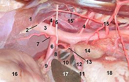
Adrenal gland
The adrenal gland ( Latin glandula adrenalis or glandula suprarenalis ) is a paired endocrine gland found in mammals , birds , reptiles and amphibians . In humans, the adrenal glands are located on the upper poles of the kidneys ; in animals that are not standing upright, they are located on the anterior kidney pole. They are subject to the hormonal control cycle and the vegetative nervous system . The adrenal gland functionally combines two different organs: The adrenal cortex , the outer part of the adrenal gland, produces steroid hormones such as cortisone and is involved in the water, mineral and sugar balance. The adrenal medulla in the inner part of the gland is part of the sympathetic nervous system and forms the "stress" hormones adrenaline and noradrenaline .
discovery
The adrenal gland was first described in 1513 by Bartolomeo Eustachi , the papal physician and anatomist, but it was not until the middle of the 19th century that they were recognized as "blood glands" ( endocrine glands ). Nobody attached any importance to it for the next two hundred years, however. It was considered to be a “gap filler” until 1855, when Thomas Addison was the first to provide a comprehensive and precise description of an adrenal gland disease with his adrenal insufficiency.
anatomy
An adrenal gland weighs about 5 to 15 grams in humans, is about 4 cm long, 4 cm thick and about 2 cm wide. In the horse it is about 8 cm long and 3 cm wide. The human right adrenal gland is triangular, the left crescent-shaped.
The adrenal glands, together with the kidneys, are surrounded by the fat capsule ( capsula adiposa ) and the kidney fascia ( fascia renalis ). Topographically, the right adrenal gland is related to the diaphragm , the right lobe of the liver , the inferior vena cava, and the right kidney. The left adrenal gland is adjacent to the left kidney, omental bursa, and stomach .
The arterial supply is ensured by three tributaries: the arteriae suprarenales superiores originate from the arteria phrenica inferior , the arteria suprarenalis media arises directly from the aorta abdominalis and the arteria suprarenalis inferior originates from the arteria renalis .
The venous drainage takes place via a central vein that emerges from the hilum of the adrenal gland. The venous blood of the left adrenal gland passes through the left suprarenal vein into the renal vein and from there into the inferior vena cava, while the blood from the right adrenal cortex passes directly into the inferior vena cava via the dextra suprarenal vein.
Histological fine structure
The adrenal glands are surrounded by a fine capsule of connective tissue and, in mammals, consist of an internal medulla and the surrounding cortex . Both parts are ontogenetically of different origins and in many lower vertebrates they form spatially separate organs. In fish they form the two separate organs, the interrenal organ and the adrenal organ . In reptiles and amphibians , both organs are attached to each other, in birds the corresponding tissue parts are interwoven.
Adrenal cortex
The adrenal cortex surrounding the medulla (Latin cortex glandulae suprarenalis ) as the outer layer ( cortex , cortex ) of the adrenal gland is of mesodermal origin and can be divided into three layers:
- Zona arcuata or glomerulosa : In the outer zone, the cells of cloven-hoofed animals and humans are arranged in a ball shape (Zona glomerulosa, from Latin glomerulum "ball"), in other mammals mostly in an arc shape (Zona arcuata, from Latin arcus "bow"). These relatively small cells produce predominantly aldosterone in response to increased levels of potassium or decreased sodium levels in the blood or decreased blood flow to the kidneys. Aldosterone is part of the renin-angiotensin-aldosterone system and regulates the concentration of potassium and sodium.
- Zona fasciculata : The middle layer is the zona fasciculata (from Latin fasciculus "strand") with relatively large cells. They are arranged like strands and rich in lipoid granules ("spongiocytes"). Sinusoidally widened capillaries lie between these cell cords . The cells mainly produce glucocorticoids such as cortisol (hydrocortisone) and corticosterone or the mineralocorticoids aldosterone and deoxycorticosterone (cortexon). Under normal circumstances, cortisol is the major glucocorticoid. The production of glucocorticoids is regulated by the adrenocorticotropic hormone (ACTH) from the pituitary gland . In addition, small amounts of sex hormones , more precisely androgens such as dehydroepiandrosterone, are synthesized.
- Zona reticularis : The zona reticularis (from Latin reticulum "network") follows the marrow with small cells arranged in a reticulate manner. They mainly form androgens, such as dehydroepiandrosterone .
All adrenal hormones are synthesized from cholesterol . The cholesterol is transported into the inner membrane of the mitochondria via a steroidogenic acute regulatory protein (StAR) . There it is converted into pregnenolone by the enzyme CYP11A . Pregnenolone can either progesterone dehydrated or 17 α hydroxylated hydroxypregnenolone. Progesterone can hydroxylation at the C 21 atom to deoxycorticosterone and converted via two further hydroxylations to aldosterone. Progesterone can be hydroxylated to 17 α -hydroxyprogesterone via hydroxylation at the C17 atom and further via deoxycortisol to cortisol.
- Memos
- The letter sequence GFR serves as a motto for the stratification of the adrenal cortex , from outside to inside: Zonae g lomerulosa, f asciculata, r eticularis. It is easy to remember because every medical professional is familiar with GFR as the abbreviation for the glomerular filtration rate .
- Similarly, the sequence salt, sugar, sex can serve for the main functions of the hormones produced in the layers: salt for aldosterone , sugar for cortisol and sex for the androgens .
- The motto for the tasks of the adrenal cortical layers "Mineral water with sugar is sexy" conveys the functions of the hormones: Mineral water for regulating the water balance (via aldosterone ), sugar for the glucocorticoid hormone cortisol , and sexy for the sex hormones.
Adrenal medulla
The adrenal medulla (Latin Medulla glandulae suprarenalis ) is located inside the adrenal gland and is the place where adrenaline and noradrenaline are produced. The adrenal medulla arises ontogenetically from the nervous system , more precisely through the migration of cells from the neural crest . These ectodermal chromaffinoblasts come from the structure of the borderline and are modified nerve cells . The marrow can also be seen as a sympathetic paraganglion .
It consists of so-called chromaffin cells (which can be easily stained with chromium salts) and multipolar ganglion cells . The chromaffin cells are formed from A cells (approx. 80% of the chromaffin cells) and N cells (also NA cells) (approx. 20%). Adrenaline is formed in A cells and noradrenaline in N cells , stored and, if required, released directly into the blood. Depending on the hormone produced , the cells are called epinephrocyti or norepinephrocyti .
The multipolar ganglion cells are part of the sympathetic nervous system . Preganglionic axons of the major and minor splanchnic nerves end at the multipolar ganglion cells .
The adrenal medulla also consists of connective tissue , blood vessels with venous plexuses and nerve fibers .
embryology
The adrenal medulla and the adrenal cortex come from different parts embryologically. The adrenal medulla originates from the ectodermal neural crest and the adrenal cortex originates from the mesodermal coelomic epithelium of the dorsal abdominal cavity, where it arises in the first embryonic month.
The retroperitoneal paraganglia, which recede after birth and lie along the abdominal aorta, fulfill the same function as the adrenal medulla before birth and release norepinephrine when there is insufficient oxygen supply. The largest retroperitoneal paraganglion is the paired Zuckerkandl organ .
Adrenal disorders
As a result of the large number of hormones formed in the adrenal gland, a variety of clinical pictures can occur in disorders.
Congenital adrenal hyperplasia is a congenital disease and is usually associated with an adrenogenital syndrome .
In the case of overactive adrenal glands ( hypercorticism , hyperadrenocorticism ), a distinction is made between two forms:
- An overproduction of aldosterone caused by a disease of the organ leads to hyperaldosteronism ( Conn syndrome ) with increased blood pressure and decreased potassium blood levels . The causes of hyperaldosteronism are either unilateral adrenal tumors ( Conn adenoma ) or bilateral enlargement of the adrenal gland ( adrenal hyperplasia ). In addition to the "classic" Conn syndrome, cases of a "normokalemic" (potassium level in the normal range) Conn syndrome are much more common and overall the most common causes of hormone-related high blood pressure . Decreased aldosterone production is known as hypoaldosteronism . It occurs especially in primary adrenal insufficiency (Addison's disease) and adrenogenital syndrome.
- An increased formation of glucocorticoids leads to hypercortisolism (also called Cushing's syndrome ). This occurs more frequently in humans, but also in dogs and horses ( equines Cushing's syndrome ). Usually the cause is an overproduction of adrenocorticotropic hormone (ACTH) in the pituitary gland , which regulates the formation of corticosteroids in the adrenal gland, more rarely a disease of the adrenal cortex itself. The resulting Cushing's syndrome manifests itself in an increased blood sugar level, trunk obesity, skin changes and bones - and muscle loss. Long-term applications with anti-inflammatory glucocorticosteroids ( e.g. prednisolone or dexamethasone ) have corresponding side effects. A reduced formation of glucocorticoids is called hypadrenocorticism (also hypocortisolism ), often combined with a general adrenal insufficiency ( Addison's disease ). This manifests itself through rapid fatigue, loss of appetite, emaciation and, in an advanced stage, through a dark, brown-yellow skin color.
Under-functions of the adrenal medulla are very rare. Overfunction of the adrenal medulla caused by tumors ( pheochromocytoma , ganglioneuroma ) can manifest itself in paroxysmal hypertension.
An acute failure of the adrenal function ( Waterhouse-Friderichsen syndrome ) can occur with septicemia , there is bleeding in the adrenal glands.
The Myelolipoma is a benign tumor that is associated mostly without clinical symptoms.
The adrenal carcinoma (also adrenal carcinoma, adrenocortical carcinoma) is a rare malignant tumor.
Adrenal hypoplasia can occur as part of a rare syndrome called IMAGE syndrome ( acronym for: Intrauterine growth retardation - metaphyseal dysplasia - congenital adrenal hypoplasia - genital anomalies).
literature
- Helga Fritsch, Wolfgang Kühnel: Pocket Atlas Anatomy . Volume 2: Internal Organs. Thieme, Stuttgart 2005, ISBN 3-13-492109-X .
- Hugo Černy, Uwe Gille: adrenal gland, glandula adrenalis s. suprarenalis. In: FV. Salomon et al. a. (Ed.): Anatomy for veterinary medicine . 2nd ext. Edition. Enke, Stuttgart 2008, ISBN 978-3-8304-1075-1 , pp. 629-631.
- Ludwig Weissbecker: Diseases of the adrenal cortex. In: Ludwig Heilmeyer (ed.): Textbook of internal medicine. Springer-Verlag, Berlin / Göttingen / Heidelberg 1955; 2nd edition ibid. 1961, pp. 1013-1025: and the same: diseases of the adrenal medulla. Ibid, pp. 1060-1063.
Web links
Individual evidence
- ^ Otto Westphal , Theodor Wieland , Heinrich Huebschmann: life regulator. Of hormones, vitamins, ferments and other active ingredients. Societäts-Verlag, Frankfurt am Main 1941 (= Frankfurter Bücher. Research and Life. Volume 1), in particular pp. 9–35 ( History of hormone research ), here: pp. 17 f.
- ↑ Sabine Schuchart: Thomas Addison had the researcher's eye . In: Deutsches Ärzteblatt . Volume 115, No. 8, February 23, 2018, p. 76.
- ↑ FeedBack What Is Adrenal Gland? Adrenal Gland Diseases. OrgansOfTheBody, accessed October 10, 2014 .
- ↑ Rothmund: Endocrine surgery. 3. Edition. Springer-Verlag, 2013, p. 391:
- ↑ a b c Ulrich Welsch, Thomas Deller, Wolfgang Kummer: Textbook Histology . 4th edition. Elsevier Urban & Fischer, Munich 2014, ISBN 978-3-437-44433-3 , pp. 443 .
- ↑ a b c Gerhard Aumüller et al .: Duale Series Anatomie , 3rd updated edition. Georg Thieme Verlag, Stuttgart 2014, ISBN 978-3-13-136043-4 , pp. 792-793.
- ^ Ulrich Welsch, Thomas Deller, Wolfgang Kummer: Textbook Histology . 4th edition. Elsevier Urban & Fischer, Munich 2014, ISBN 978-3-437-44433-3 , pp. 447 .
- ↑ IMAGE syndrome. In: Orphanet (Rare Disease Database).

