Boundary line
The sympathetic chain (lat. Sympathetic trunk ) is a chain with one another in the longitudinal direction associated ganglia of the vertebral bodies , which extends from the neck to the coccyx. It belongs to the sympathetic system .
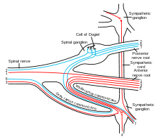
The cell bodies of the sympathetic nerve cells are located in the nucleus intermediolateralis (lateral horn) in the gray matter in the chest and lumbar region of the spinal cord . Their axons (nerve processes) leave the spinal canal and pull over the white connecting branch ( communicating branch albus ) to the respective segmental ganglion (also Paravertebralganglion , ganglion paravertebral ). There are two principal routes from the paravertebral ganglia:
- The fibers are switched to the second neuron. This is medullary and pulls over the gray connecting branch ( Ramus communicans griseus ) back to the respective spinal nerves or directly to the internal organs. For the neck, the neurons run from the stellate ganglion via the vertebral nerve along the cervical spine. From this, sympathetic fibers go to the cervical nerves.
- Some of the nerve fibers run through the paravertebral ganglia and are only switched in secondary ones , the prevertebral ganglia . These ganglia are the celiac ganglion , the superior mesenteric ganglion ( craniale ) and the inferior mesenteric ganglion ( caudale ). From here the fibers move to the internal organs. The entire sympathetic supply of the head emanates from the superior cervical ganglion.
Segments
The truncal ganglia are divided into cervical ganglia ( ganglia cervicalia ), thoracic ganglia ( ganglia thoracica ), abdominal ganglia ( ganglia lumbalia ) and cloister ganglia ( ganglia sacralia ).
There are three cervical ganglia in humans: the upper one ( ganglion cervicale superius ), the middle one ( ganglion cervicale medium ), which however is inconsistent - i.e. sometimes not present - and the lower cervical ganglion ( ganglion cervicale inferius ). The inferior cervical ganglion and the upper thoracic ganglion merge to form the larger cervicothoracic ganglion or stellate ganglion . The neck part of the border cord runs in the deep sheet of the neck fascia . It is separated from the vagus nerve by the carotid vagina and the prevertebral lamina . In veterinary anatomy, the upper and middle cervical ganglion are not included in the trunk. Here, too, the latter is not always macroscopically clear. The upper cervical ganglion, known as the cervical cranial ganglion in animals , is connected to the trunk in most mammals via the vagosympathetic trunk .
In humans there are 11 or 12 thoracic ganglia on both sides, which lie in front of the rib heads under the endothoracic fascia . The postganglionic fibers of the 2nd to 5th thoracic ganglions form the nervi cardiaci thoracici , which lead to the cardiac plexus . From the 2nd to 4th thoracic ganglion, postganglionic fibers run as nervi pulmonales thoracici to the plexus pulmonalis . From the 5th to 9th thoracic ganglion, pre- and postganglionic fibers run as the greater splanchnic nerve to the celiac plexus . From the 9th to 11th thoracic ganglion, pre- and postganglionic fibers run as the minor splanchnic nerve to the celiac plexus and the renal plexus . In some individuals there is also the splanchnic nerve that extends from the 12th thoracic ganglion to the renal plexus .
In humans, four lumbar splanchnic nerves usually originate from the lumbar ganglia of the trunk . They run with predominantly postganglionic fibers to the abdominal aortic plexus and the superior hypogastric plexus .
In humans, nervi splanchnici sacrales originate from the mostly three (up to 5) cloister ganglia of the borderline . They run with predominantly postganglionic fibers to the inferior hypogastric plexus .
The last junctional ganglion is unpaired ( ganglion impar ). It is the end of the two borderline chains and lies in front of the tailbone .
Individual evidence
- ^ Karl Zilles, Bernhard Tillmann: Anatomie . Springer, Berlin 2011, ISBN 978-3-540-69483-0 , pp. 850 .
- ^ A b Franz-Viktor Salomon: nervous system, systema nervosum . In: Franz-Viktor Salomon, Hans Geyer, Uwe Gille (Ed.): Anatomy for veterinary medicine . 2nd Edition. Enke, Stuttgart 2004, ISBN 3-8304-1007-7 , pp. 464-577 .
- ↑ Herbert Lippert: Anatomy on the living: An exercise program for medical students . Springer, Berlin 2013, ISBN 978-3-662-00661-0 , pp. 256 .
- ↑ a b c d Hans Frick, Helmut Leonhardt, Dietrich Starck: Special anatomy . tape 2 . Georg Thieme, Stuttgart 1992, ISBN 978-3-13-356904-0 , p. 606 .
