Prevertebral ganglia
The prevertebral ganglia ( ganglia praevertebralia , singular ganglion prevertebrale ) are part of the sympathetic nervous system and belong to the autonomous ganglia . They supply the abdominal and pelvic viscera.
location
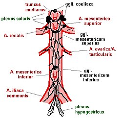
The prevertebral ganglia lie in the abdomen in front of the spine. They are grouped around the large abdominal vessels and surround the origins of the celiac trunk , the renal arteries, and the superior mesenteric artery (upper intestinal artery ).
function
Afferents
The prevertebral ganglia receive preganglionic fibers from the sympathetic neurons of the spinal cord . These fibers have their way up the paraspinal ganglia of the sympathetic trunk happened, but were there not interconnected. The switch from the first, presynaptic to the second, postsynaptic neuron only takes place in the prevertebral ganglion. The neurotransmitter of these prevertebral ganglia (preganglionic is always acetylcholine , this acts on the nicotinic acetylcholine receptors of the n 2 type) as a result, different numbers of postganglionic cells are reached ( divergence ).
Some of these preganglionic fibers, which extend from the spinal cord to the prevertebral ganglia, are particularly thick and have therefore been given their own names: Nervi splanchnici .
Afferents also draw afferents from the intestines through the prevertebral ganglia: vegetative fibers that convey pain sensations from the intestinal area to the central nervous system (CNS) via pain receptors .
Efferents
The target areas of the postganglionic fibers are the gastrointestinal tract, urinary bladder, genital organs and the walls of the surrounding blood vessels. The blood flow to the organs is regulated and bowel activity is inhibited.
coordination
In the ganglion there are connections from the viscerosensitive fibers to the efferent neurons. The ganglion therefore acts as the first central processing station outside of the central nervous system: it strengthens or inhibits afferent, triggers corresponding efferent impulses. Via these interconnections, it exerts an additional influence on the enteric nervous system .
Single ganglia
The prevertebral ganglia form a network of nerve fibers around the abdominal vessels that is difficult to see through. It is difficult to define the exact delimitation of the individual ganglia. Naming, classification and affiliations of the ganglia therefore vary from author to author.
The celiac ganglion lies around the exit of the celiac trunk from the aorta . Directly below, below the exit of the superior mesenteric artery, lies the superior mesenteric ganglion . Together with parasympathetic nerve fibers from the vagus nerve , these two ganglia form a larger nerve plexus : the solar plexus , Latin plexus solaris . Fibers from the solar plexus pull to the upper abdominal organs, including the stomach, liver and spleen.
The inferior mesenteric ganglion is located at the exit of the inferior mesenteric artery . Its postganglionic fibers participate in the superior hypogastric plexus . This nerve network consists of parasympathetic fibers from the sacral cord and sympathetic fibers and innervates the urinary and genital organs.

