Spinal nerve
A spinal nerve ( Nervus spinalis ), also known as the spinal cord (s) nerve , is the nerve assigned to one side of a certain spinal cord segment via its front and back roots . The spinal nerves are part of the peripheral nervous system . A pair of spinal nerves emerges from the spinal canal between two vertebrae . Humans have (mostly) a total of 31 paired spinal nerves.
Number of spinal nerves
The naming of the individual spinal nerves corresponds to their spinal cord segment and also follows from the spinal column sections from which they originate, their exit points. The pair of the two uppermost spinal nerves emerges directly below the occiput, i.e. above the first cervical vertebra. Since the pair of spinal nerves exiting below the seventh cervical vertebra (C8) is still assigned to the cervical area, there are eight cervical spinal nerve pairs with only seven cervical vertebrae. The spinal nerves that follow further caudally then have the same names and numbers as the vertebral body above. Overall, this generally results in humans
- Neck area: 8 pairs of cervical nerves, C1 – C8
- Chest area: 12 pairs of thoracic nerves, Th1 – Th12
- Lumbar region: 5 pairs of lumbar nerves, L1 – L5
- Sacral region: 5 sacral nerve pairs, S1 – S5
- Coccyx area: (mostly) 1 coccygeal nerve pair, Co1
Deviations are not uncommon; they can also occur in connection with malformations of the spine.
Parts of a spinal nerve
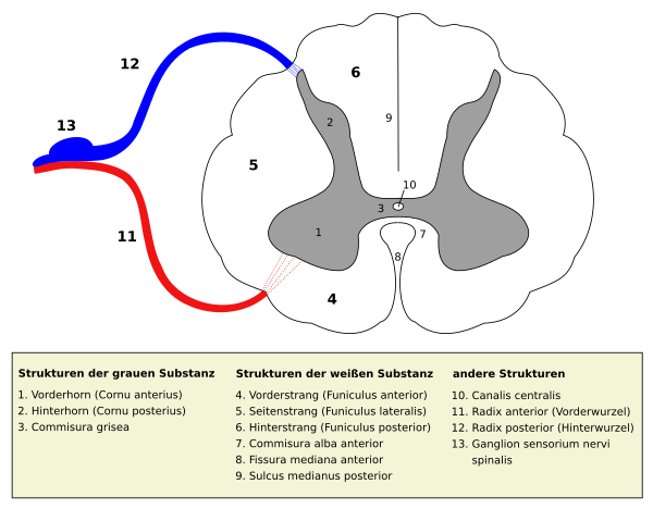
Spinal nerves are formed by the union of one efferent and one afferent nerve root , which leave the spinal cord at different points and are initially spatially separated in the spinal canal . This recognition goes to Charles Bell and François Magendie back (Bell-Magendie law) .
The afferent parts convey information from inside the body and from the body surface to the spinal cord. Their cell bodies are located in the spinal ganglion ( ganglion spinale ), which is located in the intervertebral hole. Their axons move via the dorsal root, radix posterior (in animals radix dorsalis ), into the gray matter of the spinal cord or via its white matter to the brain, where further processing takes place.
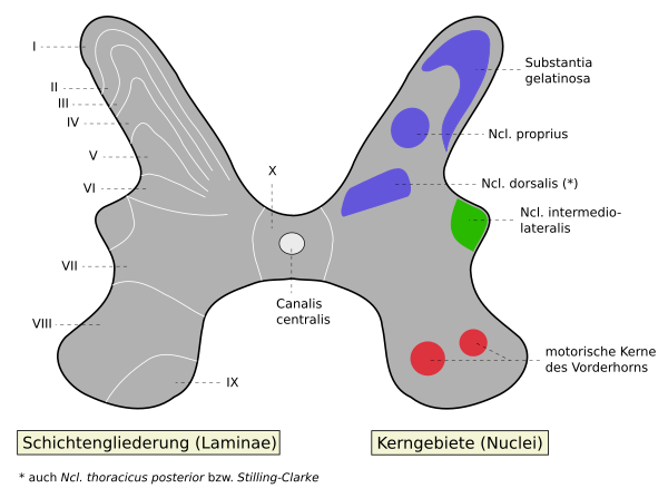
The motor efferent axons leading to a muscle are part of the motor neurons . Their somata lie in the gray matter of the spinal cord. The motor neurons of a muscle are loosely grouped in the form of a spindle-shaped nucleus in the anterior horn of the spinal cord. The efferents of a nucleus can emerge via different nerve roots. Viewed over the entire length of the spinal cord, these nuclei form what is known as the motor core column. In each segment, efferent axons emerge from the spinal cord via the anterior root, radix anterior / ventralis , and merge with the incoming afferents to form a common spinal nerve trunk ( trunk nerve spinalis ).
There are also sympathetic root cells in the chest and lumbar sections of the spinal cord . They are located in the intermediate nucleus of the gray matter and also extend in the anterior root to the spinal trunk. They then pull over a white connecting branch ( Ramus communicans albus ) to the trunk , in whose ganglia some of the fibers are switched to the so-called second neuron. The switched parts typically move back to a spinal nerve trunk in a gray connecting branch ( Ramus communicans griseus ).
There are parasympathetic root cells in the area of the cross marrow. These efferents also move to the common spinal nerve trunk. They supply the lower abdominal and pelvic viscera.
Course outside the spinal canal
The spinal trunk leaves the spinal canal via the intervertebral foramen ( foramen intervertebrale ) and then branches into several branches:
- Ramus posterior ( Ramus dorsalis ) for the care of the skin and muscles near the spine ( autochthonous back muscles ).
- Ramus anterior ( Ramus ventralis ) for supplying the skin and muscles of the back distant from the spine, the lateral and abdominal parts of the body and the extremities.
- Ramus communicans albus and Ramus communicans griseus for forwarding visceroefferent and visceroafferent information
- Ramus meningeus for the innervation of the membranes of the spinal cord
The area of skin supplied by a spinal nerve is called a dermatome , the muscles supplied as a myotome .
Plexus
Especially in the area of the origin of the limbs, the anterior / ventral rami of the spinal nerves form plexuses ( plexuses ) with their neighbors. Fibers from several spinal cord segments mix and form nerves. Each of these plexus nerves thus has parts of several spinal cord segments. Therefore, only a nerve root or a spinal nerves not occur in case of failure to complete paralysis ( paralysis ) of the muscles supplied, but only to a more or less pronounced loss of strength ( paresis ).
See also
Nervous system - motor neuron - self-reflex - axonotmesis - neurapraxia
literature
- Martin Trepel: Neuroanatomy. Structure and function. 4th revised edition. Elsevier, Urban & Fischer, Munich et al. 2008, ISBN 978-3-437-41298-1 .
- Franz-Viktor Salomon: nervous system, systema nervosum. In: Franz-Viktor Salomon, Hans Geyer, Uwe Gille (Ed.): Anatomy for veterinary medicine. Enke, Stuttgart 2004, ISBN 3-8304-1007-7 , pp. 464-577.

