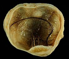
Teratoma
| Classification according to ICD-10 | |
|---|---|
| D37-D48 | Neoplasms of unsafe or unfamiliar behavior |
| ICD-10 online (WHO version 2019) | |

A teratoma (from the Greek teras " terrifying image , monster" and the suffix -om , here in the sense of "similar", hence "monstrosity"), formerly also called miraculous tumor , is an innate, often organ-like mixture of swelling , consisting of primitive, pluripotent stem cells developed. A distinction is made between mature (koätane - benign) and immature (dedifferentiated - malignant: teratocarcinoma ) forms.
The teratoma is a germ cell tumor that can therefore develop in the direction of all three germ layers . Mature forms (such as dermoid cysts ) may contain tissue such as hair or teeth that would not otherwise be found where they appeared and that did not result from metaplasia .
It is mostly in an encapsulated form that contains various differentiated types of tissue, for example skin , hair , teeth , muscle and nerve tissue . So if it contains well-differentiated tissue from all of the cotyledons it is called an adult or mature teratoma; Immature or embryonic teratomas, on the other hand, contain poorly differentiated epithelial or mesenchymal tissue. Teratomas are particularly common in the testes and ovaries .
Occurrence
Typical places of development are the ovaries (mostly benign ) or testes (mostly malignant there - teratocarcinoma ), here one assumes a poorer healing prognosis. Further localizations are: tailbone ( coccyx teratoma ), central nervous system , cervical soft tissues , mediastinum , abdominal viscera ( pancreas , intestine ), retroperitoneal space .
Teratomas make up 15–20% of benign ovarian tumors. 15% of the tumors are bilateral . More than 80% of mature teratomas occur during the reproductive phase. They are rare in children or after menopause .
pathology
Teratomas arise from primitive, pluripotent germ cells. Therefore they are counted among the germ cell tumors . The germ dispersal theory is used to explain the pathogenesis . It is assumed that germ material remains in the embryonic development and could not develop further.
histology
Teratomas usually consist of tissues from all three cotyledons , which can sometimes be arranged like an organ. It is being discussed that stem cells form tissue that is completely untypical for the environment (hair, teeth, skin - but also, to some extent, liver, kidney or heart muscle cells). The dermoid or dermoid cyst (also known as “dermoid tumor”) as a mature teratoma contains skin material (sebum glands, squamous epithelium and hair follicles) in its walls .
Histologically, in almost all cases, ectodermal tissue including cornified epidermis , sebum and sweat glands, hair follicles and neuroectodermal elements dominate. Mesodermal areas include smooth muscles, bones, teeth, cartilage, and adipose tissue.
morphology
Mature teratomas are easily recognizable macroscopically. A cystic cavity is filled with yellow sebum-like material mixed with hair. The lining of the cyst is like skin . One or more polypoid formations consisting of fatty tissue protrude into the cyst lumen (so-called head humps). Teeth, bones, cartilage, thyroid tissue or brain tissue can in some cases be observed macroscopically.
Respiratory and gastrointestinal tissue, thyroid, salivary gland and, rarely, the retina, pancreas, thymus, adrenal gland, pituitary gland, kidney, lungs, breast and prostate are derived from the endoderm . A lipogranulomatous inflammation can often be detected in the cyst wall as a reaction to the contents of the cyst. Malignant degeneration of individual tissue components occurs in only 2% of all dermoid cysts. The most common are squamous cell carcinomas or adenocarcinomas .
Teratomas, which consist of only one type of tissue, are also called monodermal teratomas. B.
- Epidermoid cyst , pure epidermal lining without skin appendages such as hair or glands
- Goiter ovarii , consisting exclusively or predominantly of thyroid tissue ; The ovarian goiter is the most common form of monodermal teratoma. It is rarely associated with an overactive thyroid or ascites . Only around five to ten percent correspond histologically to papillary thyroid carcinoma , although distant metastases are even less common.
- Carcinoid , made up of gastrointestinal or respiratory tissue
Only 3% of teratomas in women are immature teratomas with a potentially malignant course. Immature teratomas are solid or solid-cystic, have a soft, fleshy cut surface with hemorrhages and necrosis . Embryonic, mostly neuroectodermal tissue can be detected histologically . As a rule, immature tissue of the fetal type and mature tissue of the adult type from all three cotyledons are also mixed in.
Fetiform teratomas, which consist of a cyst that contains structures similar to a malformed fetus ( homunculus ), are very rare .
Symptoms
The patients are often asymptomatic depending on the location of the teratoma. Occasionally, those affected notice an increase in the circumference of the abdomen, a bulge in the lower abdomen, or complain of stomach pain.
Diagnosis

If teeth are present, the diagnosis can easily be made radiologically ( projection radiography , computed tomography CT).
therapy
The decisive factor for the treatment is whether the teratoma is benign or malignant. Benign teratomas are usually only removed surgically, immature teratomas (or teratomas in men) are also treated with chemotherapy. By cisplatin exceptionally good cure rates are -based chemotherapy, depending on the location, accessible.
Operative therapy in childhood
Complete surgical removal in sano (R0 situation) is considered to be an adequate therapy for mature teratomas; even immature teratomas of childhood can be extracranial localization according to Marina et al. adequately treated surgically.
chemotherapy
The milestone for the successful treatment of germ cell tumors was set in the 1970s with the introduction of cisplatin-based chemotherapy. Cisplatin is considered to be the most effective agent for treating germ cell tumors, although attempts have been made to replace cisplatin with carboplatin in view of its cumulative nephro- and ototoxicity .
Complications
Possible complications are twisting of the tumor with infarction , perforation, bleeding into the abdominal cavity and auto-amputation of the tumor. A sudden rupture can lead to the acute abdomen . Emptying of cyst contents can also cause granulomatous peritonitis .
particularities
Since the tumor forms stem cells - from which hair, teeth, etc. differentiate - the tumor is of great interest to developmental biologists, as it offers the possibility of obtaining stem cells.
"Growing Teratoma" Syndrome (GTS)
Microscopic foci of teratoma can grow locally and lead to compression of the surrounding structures; this observation was referred to by Logothetis in 1982 as the "growing teratoma syndrome". The clinical definition of a GTS requires
- a non-seminomatous (testicular) germ cell tumor with a history of teratomatous portion
- increased levels of the serum tumor markers AFP , bHCG and / or LDH with radiological evidence of metastasis
- the normalization of serum tumor markers after chemotherapy
- the enlargement of the metastasis (s) in normal tumor markers
- evidence of a mature teratoma in the metastasis (s).
Teratomas with malignant transformation
In rare cases, mature or immature teratomas can undergo malignant transformation (TMT). Teratomas with a malignant transformation in the traditional sense represent a well-circumscribed unit whose name refers to the malignant transformation of a somatic teratomatous component within a non- seminomatous germ cell tumor into a histology indistinguishable from a somatic malignancy . Examples of these histologically transformed cell types include
- Rhabdomyosarcoma (RMS)
- primitive neuroectodermal tumors (PNET)
- enteric adenocarcinomas
- Leukemias
See also
Web links
- Macroscopic image of a teratoma in a 16-year-old on PathoPic
- HE stained microscopic image of a mature teratoma on PathoPic
- A small brain in the ovary of a 16-year-old kurier.at from January 8, 2017
Individual evidence
- ^ The suffix -om (= Greek -ωμα) Oswald Panagl Glotta 49. Vol., 1./2. H. (1971)
- ↑ a b c d Weimann A., ea: Original examination questions with commentary GK 2. Allgemeine Pathologie , Thieme Verlag, 2002, p. 201, ISBN 3131126752 , here online
- ↑ Ulbright, 2005, Mod Pathol , 18 Suppl 2, 61-79
- ↑ Forrester and Merz, 2006, Pediatr Perinat Epidemiol , 20, 54-58
- ↑ Ng et al., 1999, Cancer, 86, 1198-1202
- ↑ Benign ovarial tumours specialist information on patient.co.uk
- ^ Riede, Schäfer: General and special pathology. 1993 Thieme, ISBN 3-13-683303-9 .
- ↑ Gobel et al., 1998, Med Pediatr Oncol, 31, 8-15
- ↑ Marina et al., 1999, J Clin Oncol , 17, 2137-2143
- ↑ Logothetis et al., 1982, Cancer, 50, 1629-1635

