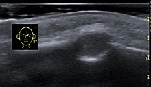
Dermoid cyst
| Classification according to ICD-10 | |
|---|---|
| D27 | Benign neoplasm of the ovary |
| D31.9 | Benign neoplasm of the eye and its appendages |
| D36.9 | Benign neoplasm in unspecified location |
| ICD-10 online (WHO version 2019) | |
The dermoid cyst (formerly also Dermoidgeschwulst ) is a cavity , which of the upper skin tissue is lined. The dermoid cyst is a teratoma .
It is a germ cell tumor, a mature teratoma made up of completely different types of tissue. Therefore, tissue structures such as muscles, cartilage, small bones, hair and also fully developed teeth can develop within the dermoid cyst.
Occurrence
Although dermoid cysts can appear anywhere, the following are comparatively common:
Ovary
In contrast to the cysts that arise during the female normal cycle and usually regress by themselves, the dermoid cyst is an embryonic malformation.
With large dermoid cysts, the risk of stem rotation (torsion) increases. Surgical removal, usually by laparoscopy, is necessary in the event of such a complication or bleeding .
Periorbital dermoid cyst
A dermoid cyst can also form in the head area ( nonodontogenic cysts ).
Together with the epidermoid cysts , dermoid cysts make up about 20 percent of the masses on the bony skull. As a rule, they already appear in infants and small children as circumscribed, initially displaceable, then no longer displaceable, smoothly delimited knots, typically at eyebrow level in the area of the entire cranial convexity.
In the differential diagnosis, an encephalocele should be considered if it is located near the root of the nose .
Spinal dermoid cyst
As part of the neural tube closure, dermoid cysts can also form on the spine, usually in the lumbar region. They are considered very rare.
Testicles
Occurrence in the male testicles indicates a high probability of degeneration .
literature
- Ursus-Nikolaus Riede (ed.): General and special pathology . Thieme, Stuttgart, ISBN 3-13-683305-8 .
- Mark K. Huntington, Robert Kruger, David W. Ohrt: Large, Complex, Benign Cystic Teratoma in an Adolescent. In: J Am Board Fam Pract. Volume 15, No. 2, 2002, pp. 164-167. ( online , PDF, 371 kB)
- M. Bettex, N. Genton, M. Stockmann (Eds.): Pediatric Surgery. Diagnostics, indication, therapy, prognosis. 2nd Edition. Thieme, Stuttgart 1982, ISBN 3-642-11330-3 .
Individual evidence
- ↑ G. Himmelfarb: On the case history of dermoid tumors of the ovary. Dermoid carcinoma ovarii dextri et Dermoid ovarii sinistri. In: Centralblatt für Gynäkologie. Volume 10, No. 35, August 28, 1886, pp. 569-571-
- ↑ "Cyst Management" http://onlinelibrary.wiley.com/doi/10.1002/uog.7610/full#bib2
- ↑ "dermoid cyst of the ovary Defined" http://www.medterms.com/script/main/art.asp?articlekey=2960
- ↑ Springer Medicine ( Memento of the original from October 22, 2013 in the Internet Archive ) Info: The archive link was inserted automatically and has not yet been checked. Please check the original and archive link according to the instructions and then remove this notice.
- ↑ Najjar et al. (2005) Dorsal Intramedullary Dermoids. Neurosurgery Review. 28: 320-325
- ↑ Aalst et al. (2009) Intraspinal Dermoid and Epidermoid Tumours: Reports of 18 Cases ad Reapproasal of the Literature. Pediatric Neurosurgery. 45: 281-290


