
Yolk sac

The yolk sac (lat. Saccus vitellinus ) is the exclusive nutritional organ of the embryos of the egg-laying vertebrates . Live birthing sharks, reptiles and mammals usually also form placentas , which in the course of further embryonic development more or less ensure complete nutrition via the maternal bloodstream. In the yolk-poor egg cells of higher mammals, the yolk sac is not only a phylogenetic relic ( rudiment ), but a necessary element in the early phase. In humans, it reaches a size of up to 5 mm and takes over some of the metabolic function of the liver before it is formed. The germ cells and stem cells for blood formation emerge from the wall of the yolk sac . With the constriction of the intestinal tube from the yolk sac and the lateral folding of the embryo, the small yolk sac lies in the chorionic cavity and is connected to the intestine via the yolk duct . The intestinal tube is separated from the roof of the yolk sac. In some mammals (e.g. horses , dogs , cats ) a real yolk sac placenta forms temporarily and the yolk sac remains as a small sac until birth.
literature
- Bertram Schnorr, Monika Kressin: Embryology of Pets. 5th edition. Enke, Stuttgart 2006, ISBN 3-8304-1061-1 .
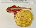
Ovoviviparous angel shark embryo with yolk sac
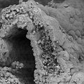
Cross section through a dog's yolk sac. Note the numerous blood cells in the wall of the yolk sac. Scanning electron micrograph

