placenta
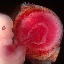
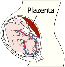
The placenta ( lat. Placenta , cakes', dt. Also placenta or "fruit cake") is an all female higher mammals ( Eutheria ), including the people and some marsupials ( Metatheria ) during gestation (or pregnancy ) evolving fabric of the wall of the uterus , which belongs to the embryonic organism, is formed by it and is interwoven with blood vessels from the mother and the embryo. The embryo (later the fetus ) is indirectly connected to the mother's bloodstream, receives nutrients and oxygen and releases waste products. After delivery, the placenta is expelled together with the egg membrane as an afterbirth . The yolk sac placenta of the basic sharks represents an analogous structure , which, like the placenta of mammals, serves to supply the embryos .
Functions
The placenta is made up of both embryonic and maternal tissue. The placenta is created when embryonic tissue grows into the lining of the uterus (endometrium or decidua). It ensures the supply of nutrients, the disposal of excretion products and the gas exchange of the embryo or fetus . The connection between the embryo and placenta is made via the umbilical cord .
In contrast to all other human organs, which only begin to function after a sufficient period of development and maturation, the placenta has to control its own growth and develop full functionality at the same time. The specific needs of the child must be met at every stage of pregnancy. In addition to caring for the child, the placenta has hormonal functions. The ability of the placenta to influence the mother's immune system in such a way that it remains functional and protects the mother from infections, but at the same time is prevented from rejecting the placenta itself and the child as foreign tissue, has not yet been researched.
construction
The human placenta is in the mature state, an about 500 to 600 grams and heavy to 20 centimeter in diameter 15 organ after implantation ( nidation ) of the blastocyst forms in the uterus. It arises from the fetal trophoblast and from the maternal uterine lining ( endometrium ). The fetal side of the placenta - i.e. the chorionic plate and umbilical cord - is covered with whitish, cloudy amniotic epithelium (see last picture). The intervillous space filled with maternal blood is located between the chorionic plate and the maternal basal plate ( decidua ) . This is divided into 15 to 20 fields, the so-called cotyledons , by connective tissue placenta septa from the basal plate . Primary villi, which carry the secondary villi, grow from the chorion into these blood-filled cotyledons. When capillaries sprout , the secondary vat becomes a tertiary vat and is therefore ready for the exchange of substances. (In the figure, the entire villus tree is designated as villus .) There is no blood exchange between the capillaries of the tertiary villi and the intervillous space due to the placental barrier (see below). The exchange of substances takes place via diffusion , facilitated diffusion, pinocytosis or is mediated via receptors. From the fourth week of pregnancy, when the child's heart begins to beat, the fruit is supplied via the placenta.
The placenta only serves as an organ for a limited time. It is characterized by the lowest content of tight connective tissue of all organs. In addition, there are no nerves pervading the placenta.
Placental barrier
One of the functions of the placenta is the placental barrier with the trophoblast-derived syncytiotrophoblast , a polynuclear layer without cell boundaries ( syncytium ). This is a passive filter membrane that separates maternal and child blood and enables or prevents the passage of various substances dissolved in the blood. The mechanisms used for this are diffusion and facilitated diffusion, active transport, diapedesis and pinocytosis . Oxygen, water, some vitamins, alcohol, poisons, drugs, and medications enter the fetus through diffusion. Glucose , amino acids and electrolytes reach the child via facilitated diffusion and active transport processes. Proteins, antibodies of the IgG type and fats are transported via pinocytosis . Viruses and bacteria can gain access to the child via diapedesis. The transmission of maternal IgG antibodies is particularly important, as the child cannot produce enough of its own antibodies until a few months after birth ( nest protection ).
Microtrauma in the placenta can cause the child's blood to pass into the maternal circulation. This is usually not dangerous unless the child is rhesus positive but the mother is negative. In such a case, the mother may become sensitized to child antigens. The mother forms antibodies of the placenta-passing IgG type against the rhesus-positive blood, which enter the fetal circulation in a subsequent pregnancy and there trigger the clinical picture of haemolyticus neonatorum .
Placenta as an endocrine organ
The placenta produces the hormone chorionic gonadotropin and from around the fourth month also the corpus luteum hormone progesterone , after the corpus luteum in the ovary stops producing. The corpus luteum hormone suppresses the menstrual bleeding and thus enables the pregnancy to continue .
In addition, the placenta forms the human placental actogen (HPL). HPL can be detected in the mother's serum from the 8th week of pregnancy and then increases continuously by up to 2 g daily until birth. The job of HPL is to develop the breasts and prepare them for lactation. It also regulates the metabolism and has anabolic effects.

Placental changes
The placenta can be very different from person to person. The shape and size of the organ vary, as does the insertion (insertion point) of the umbilical cord.
The shape variants include the placenta succenturiata (secondary placenta ), the placenta bilobata / multilobata (double / multiple lobed placenta), the placenta anularis (ring or belt-shaped placenta), the placenta fenestrata , and the placenta membranacea . A minor placenta or a lobed placenta does not interfere with fetal development.
As a consequence of a placentation disorder, forms such as the placenta accreta , the placenta increta and the placenta percreta arise . The villi grow into the myometrium through partially or completely missing decidua . The consequence is placental detachment disorders after the child is born.
Changes in position, such as the different forms of placenta previa , also occur.
afterbirth
The placenta with the membrane is born as an afterbirth shortly after the child is born.
It is often possible to have the placenta handed over. Commercial providers use it to produce so-called globules (originally homeopathic medicines). Some people bury them in the ground, usually under a tree. This custom was and is widespread in various regions of the world.
In China, the placenta was recommended against infertility and impotence in men in the 16th century. In the second half of the 20th century, placentas were also sold to the cosmetics industry . Creams made from it should be used for skin rejuvenation, there are no scientific studies on this. This practice is obsolete , among other things, because of the fear of HIV / AIDS and other infections.
The ingredients obtained from the placenta are now produced from other sources or synthetically or replaced by alternative substances.
Placentophagy
The hormone-rich placenta is sometimes also consumed by the mother to (unproven) promote regeneration and avoid postpartum depression. Critics have the suspicion that the ingestion of placenta, i.e. placentophagy, could even be harmful to the human body. This claim is based on the discovery of bacteria, lead and mercury in this very organ. The literature study by Cynthia W. Coyle et al. (Northwestern University Feinberg) from June 2015 on placentophagy in the years 1950 to 2014 comes to the conclusion that the benefits and risks still have to be researched in order to be able to make well-founded statements. Researchers at the US Centers for Disease Control and Prevention (CDC) report on a child who became infected with streptococci due to maternal placentophagy.
Stem cell extraction
It is now known that umbilical cord blood stem cells can be extracted from the placenta itself, the umbilical cord and the umbilical cord blood contained therein . In the placenta and umbilical cord, primarily mesenchymal stem cells , in the umbilical cord blood primarily hematopoietic, i.e. hematopoietic, stem cells were detected. While umbilical cord stem cells are currently only obtained for experimental and research purposes, umbilical cord blood stem cells can be routinely obtained, preserved and used medicinally. The most common area of application in 2001 was stem cell transplantation for the treatment of leukemia . However, research on the use of umbilical cord blood is still in its infancy, especially in regenerative medicine and many studies were carried out in 2013 on the cultivation of tissue, cartilage and bone parts. Cases in which umbilical cord blood is required for stem cell therapy are rare. The information from the largest stem cell bank Vita 34 can be used as evidence for this : 30 medical uses in over 200,000 storages. In addition, it is usually possible to use your own blood-forming stem cells from blood or bone marrow. Another point of criticism is the short cutting times that occur when stem cells are harvested, and are significantly shorter than the cutting times recommended by the WHO. In this context, the umbilical cord blood is often referred to as "pulsing out". The positive effects of this method have been partially proven, e.g. B. a higher amount of iron in the blood of the infant.
In animals
Most mammalian mothers - including animals that are otherwise purely vegetarian (cows and other ruminants ) - consume their own afterbirth after they have sniffed and cared for (licked dry) their newborns . Not only do they take away the tempting scent trail from the predators , they also supply themselves with vitamins and other important nutrients that they urgently need after birth.
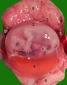
There are different types of placenta. Marsupials have an imperfect (yolk sac) placenta, which is why the gestation time is so short (8–40 days). There is also a placenta diffusa (epitheliochorial) in odd- toed ungulates and whales , a placenta zonaria (endotheliochorial) in predators , a placenta discoidalis (hemochorial) in rodents and the dry- nosed monkeys , including humans, and a placenta multiplex s. cotyledonaria or (syndesmochorial) in ruminants .
Evolution of the placenta
The placenta is a key mammalian ( evolution of mammal ) innovation from its inception to the present day. It represents a new organ that the egg-laying animals did not have before. In addition to the innovation of breast milk , the mammary gland and maternal attachment, a nutritional connection had to evolve from the egg of the embryo in the womb to the mother in order to allow the embryo to grow in the womb. That growth was a critical selective advantage. The placenta increased the chances of survival of the unborn in the time of the dinosaurs about 160 million years ago.
The gene Paternally expressed gene Peg10 was identified as an important gene for the formation of the placenta . This gene was likely encoded in the DNA of the germ cells of early mammals by a retrovirus , a viral invasion and a process similar to that observed in the recent koala with the pathogenic KoRV gene. A gene knockout of Peg10 in the mouse in the laboratory leads to a halt in the growth of the placenta and to early death of the embryo.
So Peg10 is responsible for forming the placenta. The gene suppresses the mother's immune defense and thus prevents the embryo from being rejected when the physical mother-child connection is established. Only for the further course of evolution is it assumed that the food supply of the embryo by means of the placenta was added to protect the immune system. In the course of evolution, the placenta became larger and the embryo's gestation period was extended. Longer pregnancies helped the mother to be more independent from predators, and animals began to carry live mammals to term.
The env gene of the endogenous retrovirus HERV-W is responsible for the formation of the syncytiotrophoblast . This and other ERVs involved must have acquired the common ancestors of the higher mammals, since they are absent from marsupials and monotremes (egg-laying mammals). In particular, the protein syncytin (syncytin 1 and 2) is of viral origin.
literature
- Lois Jovanovic, Genell J. Subak-Sharpe: Hormones. The medical manual for women. (Original edition: Hormones. The Woman's Answerbook. Atheneum, New York 1987) From the American by Margaret Auer, Kabel, Hamburg 1989, ISBN 3-8225-0100-X , pp. 177 f., 383 f. and more often.
Web links
- Familienplanung.de - Plazenta : The information portal of the Federal Center for Health Education
Individual evidence
- ^ Leonard Compagno , Marc Dando, Sarah Fowler: Sharks of the World. Princeton Field Guides, Princeton University Press, Princeton / Oxford 2005, ISBN 0-691-12072-2 , p. 335.
- ↑ Syncytiotrophoblast , on: DocCheck Flexikon
- ↑ Klaus Diedrich u. a. (Ed.): Gynecology and Obstetrics. 2., completely reworked. Edition. Springer, Heidelberg 2007, ISBN 978-3-540-32867-4 .
- ↑ Study overview: No, you don't have to eat your placenta. On: Spiegel Online , June 8, 2015.
- ^ Franziska Zoidl: Controversial: Lasagne with mother cake. In: Der Standard , June 19, 2015.
- ↑ Christina Hucklenbroich: Obstetrics: The consumption of the placenta - a new trend? faz.net July 15, 2015, accessed September 2, 2015.
- ↑ Cynthia W. Coyle, et al .: Placentophagy: therapeutic miracle or myth? ; In: Archives of Women's Mental Health, June 4, 2015, accessed September 2, 2015.
- ↑ Genevieve L. Buser, Sayonara Mató, Alexia Y. Zhang, Ben J. Metcalf, Bernard Beall: Notes from the Field: Late-Onset Infant Group B Streptococcus Infection Associated with Maternal Consumption of Capsules Containing Dehydrated Placenta - Oregon, 2016 . In: MMWR. Morbidity and Mortality Weekly Report . tape 66 , no. 25 , 2017, ISSN 0149-2195 , p. 677-678 , doi : 10.15585 / mmwr.mm6625a4 ( cdc.gov [accessed July 29, 2017]).
- ^ V. Lechner u. a .: Isolation of mesenchymal stem cells from the human umbilical cord as the basis for autologous stem cell therapy in pediatric surgery . German Medical Science GMS Publishing House, 2007. Doc 07dgch6655 http://www.egms.de/en/meetings/dgch2007/07dgch556.shtml
- ↑ H.-D. Peters, R. Kath: New therapeutically active monoclonal antibodies against leukemia and lymphoma . In: The oncologist . tape 7 , no. 2 , February 19, 2001, ISSN 0947-8965 , p. 196–199 , doi : 10.1007 / s007610170158 ( springer.com [accessed September 19, 2018]).
- ↑ Werner Müller: Therapy with stem cells . In: Biology in Our Time . tape 43 , no. 1 , February 2013, ISSN 0045-205X , p. 40–45 , doi : 10.1002 / biuz.201310499 ( wiley.com [accessed September 19, 2018]).
- ↑ Veronika Szentpetery: Business with fear. on: heise.de
- ↑ Susan J. McDonald, Philippa Middleton, Therese Dowswell, Peter S. Morris: Effect of timing of umbilical cord clamping of term infants on maternal and neonatal outcomes . In: Cochrane Database of Systematic Reviews . No. 7 , 2013, ISSN 1465-1858 , doi : 10.1002 / 14651858.CD004074.pub3 ( cochranelibrary.com [accessed September 19, 2018]).
- ↑ Monika Kressin, Bertram Schnorr: Embryology of Pets. 5th edition. Enke-Verlag, Stuttgart 2006, ISBN 3-8304-1061-1 .
- ↑ Ryuichi Onoa, Shin Kobayashi, Hirotaka Wagatsuma, Kohzo Aisaka, Takashi Kohda, Tomoko Kaneko-Ishino, Fumitoshi Ishino. A Retrotransposon-Derived Gene, Peg10, Is a Novel Imprinted Gene Located on Human Chromosome 7q21. Genomics, Volume 73, Issue 2, April 15, 2001, pp. 232-237.
- ↑ Ryuichi Ono, Kenji Nakamura, Kimiko Inoue, Mie Naruse, Takako Usami, Noriko Wakisaka-Saito, Toshiaki Hino, Rika Suzuki-Migishima, Narumi Ogonuki, Hiromi Miki, Takashi Kohda, Atsuo Ogura, Minesuke Yokoyko-Isokane & Fumitoshi Kino Ishino. Deletion of Peg10, an imprinted gene acquired from a retrotransposon, causes early embryonic lethality. Nature Genetics 38: 101-106 (2006).
- ↑ Nadja Podbregar: Secret Helpers - What Function Do Endogenous Retroviruses Have in Us? , on: scinexx.de from November 5, 2010
- ↑ Henning Engeln: The good side of viruses , on: Spektrum.de from April 16, 2020
- ↑ Dupressoir A, C Lavialle, Heidmann T: From ancestral infectious retroviruses to bona fide cellular genes: role of the captured syncytins in placentation . In: Placenta . 33, No. 9, September 2012, pp. 663-671. doi : 10.1016 / j.placenta.2012.05.005 . PMID 22695103 .
- ↑ Soygur B, Sati L: The role of syncytins in human reproduction and reproductive organ cancers . In: Reproduction (Cambridge, England) . 152, No. 5, 2016, pp. R167-78. doi : 10.1530 / REP-16-0031 . PMID 27486264 .
- ^ EV Koonin: Viruses and mobile elements as drivers of evolutionary transitions. In: Philosophical transactions of the Royal Society of London. Series B, Biological sciences. Volume 371, number 1701, 08 2016, p., Doi : 10.1098 / rstb.2015.0442 , PMID 27431520 , PMC 4958936 (free full text) (review).
- ↑ Mi S1, Lee X, Li X, Veldman GM, Finnerty H, Racie L, LaVallie E, Tang XY, Edouard P, Howes S, Keith JC Jr, McCoy JM .: Syncytin is a captive retroviral envelope protein involved in human placental morphogenesis . In: Nature 403 (6771) of February 17, 2000, pp. 785-789, doi: 10.1038 / 35001608 , PMID 10693809 .


