Spermatogenesis
The spermatogenesis ( ancient Greek σπέρμα sperm "seed", γένεσις génesis "Arise, Will") is the formation of sperm , that male germ cells.
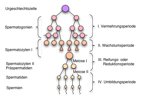
In the following, spermatogenesis in humans, i.e. in men, is discussed. Both prenatally and during puberty, spermatogonia form from the stem cells in the testes (the so-called stem spermatogonia). After puberty , spermatogonia can differentiate into first-order spermatocytes (cell enlargement). Before that, however, they divide mitotically , so that (in contrast to oogenesis ) the number of germ cells in the organism is regenerated throughout its life ( spermatocytogenesis ). Spermatogenesis can thus be divided into three stages: mitotic reproduction, meiotic maturation and finally differentiation or spermiogenesis .
stages
Multiplication
Stem spermatogonia are stored in the sexually mature testes , from which two types of spermatogonia are formed: A-spermatogonia and B-spermatogonia. The A spermatogonia emerge directly from the stem spermatogonia and mitotically divide into two daughter cells, one of which remains with the stem spermatogonia in order to ensure a constant population throughout life, while the second daughter cell continues to divide. These new cells, the B spermatogonia, go into the next phase, which is maturation.
B spermatogonia are connected by cytoplasmic processes and thus form groups that go through the following stages together and simultaneously.
maturation
The B spermatogonia migrate through the blood-testicular barrier towards the testicular tubules ( tubuli seminiferi contorti ). Now they are called 1st order spermatocytes (primary spermatocytes). These then go through the 1st meiosis ( meiosis I , haploidization ), from which two spermatocytes of the 2nd order (secondary spermatocytes) emerge. This is followed by the second meiosis ( meiosis II , equation division), from which two spermatids emerge. Four spermatids have thus arisen from one spermatocyte of the 1st order, which develop into sperm through the last step - spermiogenesis.
Spermiogenesis
In spermiogenesis, the spermatids mature into sperm. This leads to nuclear condensation, a loss of cell plasma and the formation of a kinocilium (a tail). In addition, the acrosome , which is later used to penetrate the egg cell, originates from the Golgi region. From a single, diploid spermatogonium, four haploid sperms arise through meiosis, two of which have an X chromosome and two have a Y chromosome .
Schedule
The process of spermatogenesis takes about 64 days in humans. During this time, the developing germ cells "migrate" from the base of the seminiferous tubules ( seminiferous tubules ) to the lumen of the seminiferous tubules, where the mitotic proliferation of spermatogonia about 16 days, the first meiotic division approximately 24 days and the second meiotic division only a few hours and the spermiogenesis lasts 24 days again.
Hormonal regulation
The pituitary LH ( luteinizing hormone ) controls testosterone production through a negative feedback mechanism involving the hypothalamus . The testosterone that is formed is transported into all tissues via the blood and lymph, but acts primarily in the brain and genital organs. There the testosterone has to pass the blood-testicular barrier , as it is crucial for spermatogenesis. This is made possible by the also pituitary FSH : The FSH promotes the construction of testosterone-binding proteins in the Sertoli cells , which it transports through the blood-testicular barrier.
Also important for spermatogenesis are the Leydig intermediate cells , which go through two flowering stages: during the embryonic development of the testes and LH -induced during puberty. You can now produce testosterone.
Comparison to oogenesis
The female counterpart to spermatogenesis is oogenesis . Compared to oogenesis, hydromobile compact cells are formed in spermatogenesis, which are able to actively fertilize an egg cell. In spermatogenesis four sperm are created, where the analogous steps of oogenesis only produce one egg cell . While the cytoplasm with the mitochondria is largely removed during spermiogenesis , both are retained in the oocytes. Also in men there are no pronounced cycles of hormone regulation that control oogenesis in women.
literature
- Alfred Benninghoff, Detlev Drenckhahn: Anatomy. Macroscopic anatomy, histology, embryology, cell biology. Volume 1. Cell and tissue theory, development theory, skeletal and muscular system, respiratory system, digestive system, urinary and genital system. 17th, revised edition, Elsevier / Urban & Fischer, Munich 2008, ISBN 978-3-437-42342-0 , pp. 806-807.
- Renate Lüllmann-Rauch: pocket textbook histology (= pocket textbook. ). 3rd, completely revised edition, Thieme, Stuttgart / New York 2009, ISBN 978-3-13-129243-8 , pp. 471–477.
