abdomen
The abdomen ( Latin ) or the belly is the area of the trunk between the chest and pelvis in anatomical terminology . The associated adjective is abdominal .
Regions of the abdomen
The upper limit of the abdomen can be at the level of the breastbone schedule lace, the lower limit on the inguinal ligament . The area above the navel framed by ribs is the upper abdomen . The area without bony elements is the mid-abdomen. The lower abdomen (also lower abdomen ) is in turn surrounded by the pelvis . If you also subdivide in the longitudinal direction along the outer edges of the straight abdominal muscle , you get the stomach region , the umbilical region and the pubic region in the middle from top to bottom and the rib region, the outer region and the groin region on the outside .
Surface of the abdomen
On the front, the abdominal wall , occurs depending on the training condition more or less clearly the both sides just created abdominal muscle ( musculus rectus abdominis of up to the level of the navel by three transversely extending narrow chord plates) produced, - inter cesarean sections tendineae - is interrupted. The navel is roughly in the middle of the abdomen. The upper part of the abdomen is bordered by the lowest ribs that run towards the tip of the sternum (the sword extension ). In the lower area outside is the upper edge of the pelvis (crista iliaca). In most people, the muscle relief is covered by a thick layer of fat, the subcutaneous fat tissue is the largest fat depot , especially in men . The sternum is always palpable above and the anterior upper tip of the upper edge of the pelvis (iliac spine, spina iliaca) below, from which the skin appears constricted along the inguinal ligament as the border to the leg . In the pubic area , the upper edge of the pubic bone can be felt with the symphysis .
Abdominal cavity

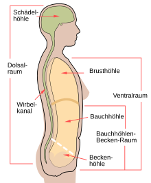
The cavity of the abdomen is called the abdominal cavity (also abdominal cavity or in Latin cavitas abdominalis ). It is limited upwards (cranially) by the diaphragm , downwards (caudally) by the hipbone with the superimposed muscles and by the pelvic floor , laterally and forwards (ventrally) by the anterior abdominal wall and to the rear (dorsal) by the lumbar spine, the sacrum and the deep abdominal muscles .
The abdominal cavity through which is lined parietal peritoneum lining as a wall sheet of the abdominal cavity the outer walls and itself as a visceral peritoneum ( visceral leaf) in the abdominal and pelvic organs continues and these covers to a large extent.
The abdominal cavity is divided by the peritoneum into the surrounding peritoneal cavity (Cavitas peritonealis) , the retroperitoneal space (Spatium retroperitonealis) lying dorsally from it and the subperitoneal space lying caudally (Spatium subperitoneale) .
Pelvic cavity (Cavitas pelvis) is the name of the section of the abdominal cavity lying caudal to the pelvic entrance level. It contains parts of all three previously mentioned sections.
"Abnormal content" of the abdominal cavity is an increased fluid content ( ascites ) , blood ( hemascus ) , lymph (chylascus) and gas ( pneumoperitoneum ) . A perforation of the stomach wall allows stomach contents, a perforation of the intestine intestinal contents or feces into the abdominal cavity.
Separate rooms
The abdominal cavity is divided into the upper and lower abdomen by the mesentery of the transverse bowel (the mesocolon of the transverse colon). Mesocolon, mesentery and other peritoneum duplicates divide the abdominal cavity in humans into several gap-shaped spaces, so-called peritoneum niches or pockets:
In the upper abdomen:
- The subphrenic recess (right and left subphrenic gap) are separated from each other by the falciform ligament and lie between the front of the liver and the underside of the diaphragm.
- The recessus or sulci paracolici represent the continuation of these cleft formations downwards and are bounded on the right by the lateral abdominal wall and the ascending, on the left by the lateral abdominal wall and the descending part of the large intestine.
- The subhepatic recess is bounded above by the underside of the liver and below by the transverse portion of the large intestine, the stomach and the small network of the lesser omentum .
- The omental bursa as the largest peritoneum niche in the abdominal cavity.
- The recessus morisoni is bounded in front by the rear of the right lobe of the liver and in the rear by the right kidney located retroperitoneally.
In the lower abdomen:
- The right mesenteric colic gap ( Spatium mesenterico-colicum dexter ) is reached via the insertion of the mesentery, the so-called mesenteric root .
- The left mesenteric colic gap ( Spatium mesenterico-colicum sinister ) is reached under the attachment .
- The retrocaecal recess lies behind the appendix and is accessible from its underside.
- The intersigmoid recess under the sigmoid colon corresponds to the retroperitoneal course of the left ureter .
Abdominal organs
Various organs are found in the abdominal cavity. If the organs are covered by the peritoneum, they are called intraperitoneal , if they are behind they are called retroperitoneal . They can also be divided into upper and lower abdominal organs. Upper abdominal organs include the liver , gallbladder , stomach , duodenum , pancreas, and spleen . The lower abdominal organs include the small intestine (excluding the duodenum) and the large intestine (including the transverse colon, excluding the rectum ). The surface of the abdominal organs or their peritoneal lining is covered by a thin serous film of liquid ( liquor peritoenei ), which ensures that the abdominal and pelvic organs can be moved relative to one another.
topography
At the front of the spine is the borderline , which contains the vegetative nerve fibers for the organs . To the left in front of the spine is the aorta , the main artery from which all other arteries branch off. To the right in front of the spine is the inferior vena cava , the main vein .
The liver lies in the right and middle upper abdomen and nestles under the right diaphragmatic dome. To the left of it is the stomach with its curve open towards the liver . Both organs are largely covered by the rib cage .
The gall bladder is hidden under the liver, and the coffee bean-shaped spleen is to the left behind the stomach.
The stomach continues into the small intestine , at the beginning of which the elongated pancreas has grown, the tip of which points towards the spleen behind the stomach.
The small intestine consists of a random tangle of intestinal loops, which fills most of the middle and lower abdomen. It is divided into three sections: duodenum (duodenum), jejunum (empty intestine) and ileum (ileum or hip intestine ). The ileum opens into the large intestine in the right lower abdomen .
The large intestine (colon) has a rudimentary , blind end at its beginning downwards : the caecum (appendix) with the appendix vermiformis ( appendix ), the position of which can be very different. It continues upwards as the ascending colon and extends up to the liver. It is suspended there by the flexura coli dexter . As a transverse colon ( Colon transversum ), the large intestine then pulls in front of the stomach and the loops of the small intestine to the left side where it is suspended from the flexura coli sinister . From there it descends as the descending colon in the direction of the anus . It visually forms a kind of frame around the loops of the small intestine.
The kidneys are located in the retroperitoneal space on both sides of the spine . The upper pole is at the level of the 12th thoracic vertebra, the lower pole at the level of the 3rd lumbar vertebra. The right kidney is about half a vertebral body lower than the left because of the liver.
embryology
In the embryo , all organs are initially located behind the peritoneum , the peritoneum, i.e. retroperitoneally . The kidneys with the adrenal glands also stay there. The digestive tract (stomach, small intestine, large intestine; see gastrointestinal tract ) then turns forward into the peritoneal space ( i.e. intraperitoneally ), with certain sections of the intestine then attaching themselves to the peritoneum (secondary retroperitoneal). For this reason, all sections of the digestive tract are connected to the abdominal wall via a fold in the peritoneum, a so-called mesentery , at the back - and in the upper abdomen also at the front . The liver is an ingrowth in the anterior mesentery of the stomach, the mesogastrium ; the spleen and pancreas are ingrowths in its posterior mesentery (both cf. mesentery ). Because of the twisting of the stomach , the liver has come to lie to the right of the stomach; the pancreas and the spleen on the left behind.
All conduction pathways reach their target organs, provided they are within the peritoneal space, via such a mesentery.
The kidneys, the spleen and the liver each have a "gateway", a so-called hilum , i.e. an indentation on the inward-facing side through which all conduction pathways reach or leave the organ.
stomach pain
The cause of abdominal pain disorders come from organs (such as pancreas or gallbladder) of the abdominal cavity and (intra-abdominal) Infections (eg peritonitis , biliary tract or intra-abdominal abscesses ) in the abdominal cavity, diseases that take place outside of the abdominal cavity, but also discomfort caused by mental illnesses are raised in question. Organic pain can usually only be roughly specified as upper abdominal pain, pain in the middle abdomen, and lower abdominal pain. A testicular pain can be projected in the abdominal area (eg. As testicular torsion ). A heart attack can also rarely manifest itself as pain in the upper abdomen (see also head zone ). A pain on the skin or in the abdominal wall ( muscle tension ), on the other hand, can be localized more precisely.
Examination of the abdomen
The abdominal examination is initially carried out by inquiring about the symptoms ( anamnesis ), inspection (looking at), auscultation (listening), percussion (tapping), palpation (palpation) and a blood test . Depending on the result of this examination, imaging procedures such as conventional X-rays , sonography , computed tomography (CT) or MRI can provide an insight into the inner workings.
When inspecting the abdomen can be found among other things
- Retraction of the abdomen (passive or - in the case of the scabbard - active retraction of the abdominal wall)
- Bulging of the abdomen, caused by
- the fat distribution
- Ascites (accumulation of fluid in the abdomen)
- Meteorism (abdominal distension)
- the shape of the pelvis
- Hairiness and pigmentation
- abnormal vascularization of the abdominal wall (especially blood vessel dilation)
literature
- Tina Ebbing: Center of the body. A cultural history of the stomach since early modern times. Campus, Frankfurt am Main / New York 2008. Zugl .: Dissertation, University of Münster 2007, ISBN 978-3-593-38733-8 ( review at www.sehepunkte.de ).
- Abdominal cavity
- Herbert Lippert: Textbook anatomy. 7th edition. Elsevier, Munich 2003, ISBN 978-3-437-42362-8 , p. 278.
- Helga Fritsch, Wolfgang Kühnel: Pocket Atlas Anatomy - Volume 2 Inner Organs. 9th edition. Georg Thieme Verlag, Stuttgart 2005, ISBN 3-13-492109-X , p. 182.
- Examination of the abdomen
- Herrmann S. Füeßl, Martin Middeke: Anamnesis and clinical examination. 3. Edition. Georg Thieme Verlag, Stuttgart 2005, ISBN 3-13-126883-2 .
Web links
- Classification of the abdominal regions ( memento from February 24, 2009 in the Internet Archive )
- Organs in the abdomen (private side)
Individual evidence
- ^ Pschyrembel Clinical Dictionary. 259th edition. Walter de Gruyter GmbH & Co, Berlin 2001, ISBN 3-11-016522-8 , pp. 808, 1446.
- ^ Marianne Abele-Horn: Antimicrobial Therapy. Decision support for the treatment and prophylaxis of infectious diseases. With the collaboration of Werner Heinz, Hartwig Klinker, Johann Schurz and August Stich, 2nd, revised and expanded edition. Peter Wiehl, Marburg 2009, ISBN 978-3-927219-14-4 , pp. 119-129 ( intra-abdominal infections ).
- ^ Klaus Holldack, Klaus Gahl: Auscultation and percussion. Inspection and palpation. Thieme, Stuttgart 1955; 10th, revised edition, ibid. 1986, ISBN 3-13-352410-0 , pp. 230-238.

