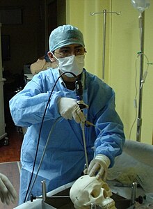
endoscope
An endoscope (formed from ancient Greek ἔνδον éndon , German 'inside' and ancient Greek σκοπεῖν skopein 'observe') is a device with which the interior of organisms or technical cavities can be examined and manipulated. Known as the application of the endoscope endoscopy was originally for human medical diagnosis developed. Today it is also used for minimally invasive surgical interventions on humans and animals as well as in technology for the visual inspection of cavities that are difficult to access.
Basic types and working diameter
The endoscopes include the rigid and flexible endoscopes and their subspecies. Different names are often used for endoscopes depending on the manufacturer, e.g. B. for rigid endoscopes: borescopes or boroscopes, technoscopes, autoscopes, intrascopes; for flexible fiber optic endoscopes etc. a. Fiberscopes or flexoscopes.
Common working diameters of rigid borescopes are 1.6 to 19 mm. Semi-rigid borescopes (also called elastic or semi-flexible) are available from 1.0 mm, flexible endoscopes from 0.3 to 15 mm and video endoscopes from 3.8 to 12 mm.
Rigid endoscopes
A rigid endoscope ( Eng./techn. Rigid Borescope ) transmits the image information of the object or space to be examined through a lens system inside the endoscope shaft to the eyepiece . Examples are the technical borescope and, in the medical field, the arthroscope and cystoscope .
The rod lens system developed by Harold H. Hopkins is widespread . Here the light is passed through rod lenses made of quartz glass and refracted by air lenses between the rods. This very bright construction enables smaller lens diameters. Most current endoscopes offer the possibility of setting the image to the optimum sharpness even for those who wear glasses by means of a focusing ring near the eyepiece. The light from the light source required for the examination / inspection is transported to the tip of the endoscope via the connected light guide, also inside the shaft through glass fiber bundles. The price of a rigid endoscope depends on the quality of the lenses used, the viewing / viewing angles of the lens and the working length or working diameter . On average, this is an amount in the rather lower four-digit euro range.
Rigid endoscopes with swivel prism reflecting on the objective side can look to the side in cavities. By rotating the endoscope in its main axis and pivoting the prism which deflects the direction of view therefrom, a larger solid angle in the cavity can be viewed by scanning. A small, polished metal mirror, which is connected to the endoscope lens head with a flexible wire and a push-on sleeve, does a similar thing.
Flexible endoscopes
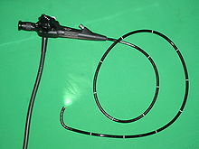
With a flexible endoscope (or flexoscope or Fiberscope , the naming is partly manufacturer- dependent ), the image and light are transmitted via fiber optic bundles. Examples are the technical flexoscope and, in the medical field, the gastroscope , colonoscope , bronchoscope and arthroscope . The cardiac catheter also belongs to the class of endoscopes.
From a practicable diameter, fiberscopes / videoscopes are also available with exchangeable instead of fixed lenses ( front / side or backward ) as well as working channels for inserting micromechanical devices (small pliers or grippers) into the examination or inspection room. Flexible glass fiber endoscopes (fiberscopes) and video endoscopes (videoscopes) usually have a device tip that can be remotely controlled via built-in Bowden cables . Depending on the model and diameter, this can be angled up to 180 ° on 2 ( up-down ) or 4 sides ( up-down and right-left ). The length of this bendable tip can be between 30 and 70 mm depending on the diameter. A mechanism is built into the handle of the device, which acts on the Bowden cables via tilt bars or handwheels and enables this movement of the tip.
See also: Medical Endoscopy and Micromechanics
Video endoscopes
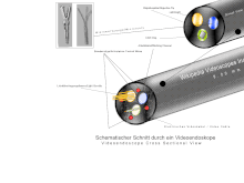
The youngest subtype of flexible endoscopes are the video endoscopes, often also called videoscopes (English videoscope or video sample), whereby the name depends on the manufacturer. Video endoscopes open a new chapter in modern endoscopy as they use digital technology to generate and transmit images . A chip attached to the lens of the video endoscope (see also: digital camera ) generates an image of the examination subject. With the CMOS chip, the image signal is digitized on the video chip so that the image transmitted to the video processor can be influenced less by electromagnetic interference than with endoscopes with a CCD chip, where the analog signal from the chip only occurs outside the endoscope in the so-called Video processor is digitized for further processing. A video processor prepares the image information and combines it with examination data and patient information before the images or video sequences are displayed on the viewing screen or saved on a storage medium. A transfer to the clinic network can also take place from here. Just like fiber endoscopes, video endoscopes also have a remotely controllable bendable device tip that can be moved in 2 or 4 directions depending on the intended use. It is controlled mechanically or electronically. The mechanical control takes place via a small gear unit using rocker arms or rotary wheels. Instead of the mechanics, some videoscopes have built in small electric motors that control the Bowden cables via a joystick.
Video endoscopes generally have a resolution many times higher than fiber endoscopes. The image quality is significantly influenced by the quality of the lens system and the video chip as well as the lighting of the examination area and the post-processing of the image signal in the video processor. The last crucial link in the chain of image reproduction is the viewing screen. For many years the CCD chip offered the best video quality. Due to the compact design, chips of this type are still mostly used in video endoscopes today. The latest CMOS chips enable higher frame rates than CCDs with higher resolution at the same time, so that the first video endoscopes today achieve a display in 1080p. Devices that use LEDs in the tip of the device for illumination do not currently achieve the same image quality as those that illuminate with light guides due to the lower light intensity. Battery-operated LED light sources, which are attached directly to the endoscope, are only a special solution for exceptional situations due to the even weaker illumination. The current development in lighting technology is the multi-LED light source. The light from several LEDs is here in an external light source bundled and, as with the xenon light source, emitted via one or more light guides onto the examination area. This technology enables illumination that meets at least the same requirements as xenon light, while at the same time the lamp has a much longer service life and significantly lower energy consumption.
Manufacturers of digital video endoscopes include Olympus, Pentax Medical, Fujifilm and Karl Storz.
Basic components
A simple endoscope set includes:
- Light source
- Light guide
- Endoscope (with image guide)
As a rule, individual components from different manufacturers cannot be easily combined. A light guide or endoscope from one manufacturer, for example, cannot easily be operated on a light source from another manufacturer. Well-known manufacturers offer suitable adapters on request. Various support arm systems are offered by the industry to facilitate practical work with endoscopes .
Light sources
In particular, the use of digital image transmission techniques ( video endoscopy ) using CCD chips made the use of expensive xenon lamps necessary. Their light intensity is excellent, but their service life is largely determined by the respective on / off cycles. The following applies: the more cycles, the shorter the service life.
Light sources such as xenon lamps develop an enormous amount of heat during operation, which is largely caused by the infrared component of the light source spectrum . It must therefore be prevented that the IR component reaches the light exit of the endoscope. Modern light sources can therefore be regulated in terms of light intensity , cooled by a fan and the infrared radiation is largely removed from the light spectrum by dichroic concave mirrors and (additionally) by heat protection filters in front of the light guide. These systems are called cold light sources and the light sources are called cold light mirror lamps . Another development that is advantageous due to the low power / cooling requirement are devices with light emitting diodes (LED) as a light source. However, the light output of LEDs cannot yet compete with that of xenon lamps. Nevertheless, this technology opens up new areas of application and offers an interesting alternative, especially for light sources in battery operation.
Light guide
Glass fibers are mainly used for endoscopic light guides. But there are also light guides that can guide the light using a gel as a transport medium. Gel light guides, also known as liquid light guides, offer a higher light output, which is particularly advantageous for large rooms and digital endoscopy in general. Liquid light guides are usually better suited for the transmission of UV light than glass fibers. Gel / liquid light guides are a little more unwieldy to use and not as flexible as well as a little more expensive than glass fiber light guides. Without a connected light guide you can see an image through the endoscope, but this is too dark to achieve usable results in closed rooms.
Image conductor
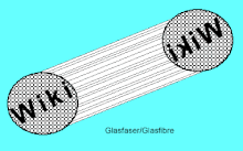
Image guides are made up of many thousands of individual glass fibers with a diameter of 7 to 10 µm. This corresponds to a resolution of 3,000 to 42,000 or 75 × 45 to 240 × 180 picture elements (pixels), depending on the diameter. Brightness and color information can be transmitted per fiber. The moiré effect caused by the superposition of the fiber grid with the CCD grid can reduce the quality of the image, which is why more and more videoscopes and video endoscopes with built-in CCD chips are used at the distal end.
measuring technology
In the field of measurement technology , various manufacturers offer measurement methods. Each measurement technology has advantages and disadvantages for the respective purpose. In technical endoscopy in particular, measuring systems are used in many areas of application, some of which today can deliver surprisingly accurate results. Areas of application for this are aircraft turbines or power plant areas. There are currently four non-contact measurement methods used in video endoscopes, some of which are also used in rigid endoscopes:
- Shadow / video image measurement ("Shadow")
- "2-point laser measurement" - naming z. Partly manufacturer-dependent
These types of measurement only provide an exact measurement if the measuring endoscope is aligned vertically on the surface to be measured.
The measurement methods are much more precise:
- Comparative measurement ("stereo")
- Laser measurement ("Multipoint")
Current research deals with the possibility of capturing 3D data endoscopically. For this purpose, the approach of fringe projection is usually used as a variant of flat triangulation . Depending on the optics used, technical internal geometries can be recorded and evaluated with resolutions in the lower µm range.
As with any measuring device, the measuring accuracy of an endoscope measuring system depends crucially on the training and experience of the user. A fully equipped, measurable video endoscope can cost a high five-digit euro amount.
optics
Regularities
In connection with the working diameter of an endoscope, the following applies:
The larger the diameter, the brighter and wider the image.
According to the laws of optics , there is also the following relationship between viewing angle and magnification factor:
Large viewing angle = low magnification (wide angle in photography )
Small viewing angle = high magnification (telephoto lens in photography )
The following applies in relation to the magnification and the distance between the lens and the object to be examined:
The magnification factor describes the object size in the image relative to the real object size
and further:
The magnification factor is inversely proportional to the distance: Objective / object ( depending on other factors )
Lenses
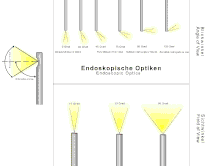
| Viewing angle in degrees ° | Direction of view / designation |
|---|---|
| 0 | Straight-ahead ( oblique or direct view ) |
| 30-80 | Forward-looking ( fore or oblique front view ) |
| 90 | Side view ( right angle or lateral ) |
| 110-120 | Looking back ( retrograde or rear ) |
Labelling
Endoscopes are marked with a key of their characteristics, this can usually be found on the shaft or the handle as an engraving. The following applies:
| Working diameter · viewing angle · viewing angle |
An endoscope with the following information: 6-70-67 would therefore have the data:
Working diameter = 6.00 mm, viewing angle = 70 °, viewing angle = 67 °.
A rather magnifying endoscope with a forward looking lens.
Timetable
- 1806 - Philipp Bozzini (city doctor in Frankfurt) constructs and describes for the first time a rigid medical endoscope and sends this "light guide" operated by candlelight to the Medical University of Vienna for assessment; there it is tried out on corpses and assessed positively.
The original Bozzini endoscope was thought to be lost after the Second World War , but was found again in the USA and returned to the Vienna Institute for the History of Medicine in 2001 via the “International Nitze-Leiter Research Association for Endoscopy”. There is no evidence of its use at the time. - 1850/51 - Hermann von Helmholtz develops the ophthalmoscope and uses it in practice
- 1855 - Further development of the “Bozzini” endoscope by the French surgeon Antonin Jean Désormeaux . ( He replaced the candle Bozzini used as a light source with a gas arc lamp. )
- 1865/1867 Julius Bruck developed the stomatoscope for x-raying the teeth and the urethroscope for x-raying the bladder with galvanic incandescent light .
- 1879 - The Dresden doctor Maximilian Nitze presents his " cystoscope " , which was made with the help of the Viennese craftsman Josef Leiter
- 1881 - Johann von Mikulicz founded esophagoscopy and gastroscopy
- 1902 - D. von Ott makes a douglass copy for the first time to inspect the Douglas space with a cystoscope.
- 1912 - urological cystoscope by Otto Ringleb
- 1958 - Development of the first flexible endoscope ( flexoscope ) by Basil Hirschowitz
- 1967 - Kurt Semm founded modern endoscopy as a gynecologist
- 1971 - Endoscopic ablation of colon polyps with a flexible instrument on March 13, 1971 at the University of Erlangen .
- 1976 - Development of the first disinfection device for flexible endoscopes by Siegfried Ernst Miederer and working group at the University of Bonn.
- 2000 - Introduction of capsule endoscopy into practice
Endoscopy pioneers are Olympus , Karl Storz and Richard Wolf GmbH .
application areas
General
Endoscopy has a wide range of uses. Endoscopes are used in medicine as well as in the technical field (technical endoscopy).
Technical endoscopy
Endoscopes are used in a variety of ways in the technical field today and are an important component of "non-destructive testing" (NDT - Non Destructive Testing).
In addition to the endoscopes, other methods such as B. the X-ray, eddy current, ultrasonic method or microscopy in non-destructive testing application. All of the methods and procedures mentioned are used to check the quality or wear and tear of components.
The above-mentioned component inspection is usually a purely visual inspection. Endoscopes are versatile due to their structural properties. For this reason, they can be used for a variety of examinations. In the 1980s, documentation of the images obtained was only possible under difficult conditions (e.g. using a reflex camera with special filter sets and long exposure times), but documentation of the images obtained is no longer a problem today. Because of this change there is an almost infinite number of test options. Here are some application examples:
- Archeology - this is how z. B. Ötzi examined endoscopically
- Architecture - Endoscopic photography in architecture has been used since 1954.
- Police and Customs - for checking the location of unsafe rooms and investigating possible hiding places for contraband or narcotics .
- Secret services or espionage
- Military and arms industry
- Investigation of musical instruments
- Rescuing buried victims in disasters
- Investigation of animal structures (e.g. mole passages or bird nests)
- Building protection - checking the insulation of old buildings / checking for pest infestation in wooden buildings / researching the cause of water damage
- Monument Preservation - Large monuments are often hollow and can be checked for any corrosive processes using endoscopy
- Automotive industry - Here the endoscope is mainly used to check cavity seals and motors (wear).
- Ship industry (engines)
- Industrial plants ( power plants , pipe welds , wind power gears )
- Quality assurance of various components up to circuit boards
- in the sanitary area to examine defective lines
- aviation
History of technical endoscopy
In aviation, endoscopy has been used for the maintenance of aircraft engines , for example, since the 1950s . With the help of the working duct and micro tools, minor repairs can also be carried out on engine blades. The term boroscopy (from English Borescope; bore " Bohrloch / Bohr") was established in this area . The English version of the flexoscope is a flexiscope or flexoscope. Endoscopy (Engl. Endoscopy ) is the generic term is for this technology.
Modern developments
In the course of increasing material and quality requirements, industrial components are more and more frequently subjected to optical series testing. Technical endoscopes are used as an aid for inaccessible surfaces. For internal testing of cylindrical objects, e.g. B. from a hydraulic cylinder, these are endoscopes with a side view, d. H. the optical axis is deflected by a mirror or prism (comparable to a periscope). For complete coverage of the surface, the object and the endoscope must be linearly displaced and rotated relative to one another. In order to avoid the time-consuming rotational movement, attempts were made to replace the deflecting mirrors with conical mirrors that were placed with the tip on the endoscope. These constructions were mechanically, optically and in practical application unsatisfactory and have not become established. In endoscopy, deflecting prisms are generally preferred over mirrors. Mirrors are very sensitive to dust or dirt and significantly affect the picture. If you virtually rotate such a prism around the optical axis of the endoscope, a simple panoramic prism is created.
In 1985, a straight-ahead endoscope was fitted with a special prism at the front for the first time. It was a glass ball into which a cone was cut from the front, which was optically mirrored. Using this “conical mirror”, the surface of a segment could now be viewed all around at a glance. The requirement at that time was 100% internal inspection of car master brake cylinders. These parts, with their honed inner surfaces, had to be flawless and without voids and scratches. Since it is an important safety part on the car, a 100% inspection was inevitable. Over the years this technology has been refined and adapted to other applications and requirements. If you grind z. B. instead of the cone, insert a radius into the glass ball, then you can even look back and do that all around. These panoramic prisms, which provide a considerably better image than mirrors, have been continuously developed and now meet the highest demands and are even suitable for automatic image processing.
Special optics
Panoramic endoscopes
The panoramic endoscope is a special technical endoscope (English borescope for industrial endoscope, in contrast to the medical endoscope) for the inspection of cylindrical cavities. An endoscope normally produces a round, disk-shaped image, whereas a panoramic endoscope produces a ring-shaped image, i.e. H. there is no image information in the center of the exit pupil. This fundamentally distinguishes the panoramic endoscope from the fisheye lens , in which the essential image information is located in the center of the exit pupil. However, a fisheye lens mainly looks forward, the panoramic prism more to the side, but all around (see illustration).
Image generation and effect
The panoramic prism consists of several spherical glass surfaces joined together. It collects the entire 360 ° image information from a length of the cylinder. The length of the section depends on the angle of view ( field of view , FOV ) of the panoramic prism and the distance between the surface and the endoscope. When viewing directly or using a matrix camera customary in the industry, the image appears radially distorted. The distortion can either be eliminated by downstream image processing or by using a ring-shaped line camera, the stripe images of which, when joined together, provide a distortion-free development of the inner wall of the cylinder.
Medical endoscopy
Medical endoscopes have to study the stomach - intestinal tract , the lungs and the uterus revolutionized. Even the draining tear ducts can be examined endoscopically.
The oldest and simplest endoscopes still in use consist of a rigid tube through which the necessary light is reflected and through which one can see with the naked eye. That is why one speaks of “mirroring” in the vernacular. The longer devices were also equipped with lenses in a tube at the front end and for the first time enabled small passive movements.
An initial further development consisted in bringing light generated remotely to the tip of the tube using fiber optic bundles. The next development step was to also transfer the image information to the examiner's eye via flexible, ordered glass fiber bundles , the image guides . Only then did the endoscope become really flexible. Since then, the device has been actively controlled via four integrated Bowden cables .
In addition to the components described under Basic , a medical endoscopy unit includes :
- imperative
- an air insufflator or a gas pump for the dosed inflation of hollow organs or body cavities (abdominal cavity), where the walls would otherwise fall on the optics or details would be hidden in folds.
In the simplest case, this is a rubber balloon with a valve (for rectoscopy, see below) that is operated by hand. For flexible endoscopies (gastroscopy, for example) a pressure-limited pump is used and the endoscopist blows in the air using finger valves. In the abdominal cavity mirror , however, one uses volume- and pressure-limited controlled machines and to avoid air embolism is CO 2 gas in place of air is blown. - an irrigator : in the simplest case a syringe or infusion bottle filled with saline solution
- a suction pump for mucus and other unwanted liquid contents of the hollow organs
- as required
- a coagulator to stop bleeding
-
flexible tools . They are brought in through working channels.
- Gripping or cutting tools for the purpose of obtaining tissue samples
- Cannulas for injection
- Wire electrodes for coagulation with electric current.
Nowadays, especially under stationary conditions, the image is no longer viewed directly with the eye (neither on the rigid tube endoscope nor on the eyepiece of the flexible endoscope), but on one or more modern monitors that falsify the color information as little as possible, and the Enable work and teaching (kibitzing) in daylight without any loss of quality. This also opens up the possibility of recording on video carriers or transmission to lecture halls.
An interesting recent development is the " endoscopic pill " or capsule endoscopy : a mini camera, which is taken orally in the form of a pill and transported through the digestive tract by natural peristalsis , takes pictures of the intestine in a continuous series . The capsule is designed for one-time use. This technique as well as the evaluation are complex, but extremely helpful in the case of hidden bleeding or small tumors in the small intestine as a " last resort ". A simultaneous therapeutic intervention as with the other endoscopic methods is currently not possible.
The disinfection of the flexible devices, which are heat-sensitive and therefore not accessible to simple methods, is of great importance . Today, modern disinfection devices guarantee that endoscopes are free of germs. The first disinfection device was developed in 1976 by a working group led by SE Miederer.
Preparation in medical endoscopy
In most endoscopic examinations, the person concerned is given premedication to make things easier, i.e. a sedative such as midazolam or the anesthetic propofol is given.
Medical endoscopic examination and treatment methods
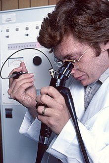
- Reflection on the gastrointestinal tract
- Esophagus ( esophagoscopy )
- Stomach ( gastroscopy )
- Creation of a gastric fistula using an endoscope ( percutaneous endoscopic gastrostomy (PEG))
- Salivary glands ( sialendoscopy )
- Duodenum ( duodenoscopy or gastroduodenoscopy )
- alone or
- with x-ray of bile ducts and pancreatic duct ( ERCP )
- Biliary tract ( cholangioscopy )
- Small bowel endoscopy (enteroscopy and capsule endoscopy )
- Colonoscopy ( proctoscopy , rectoscopy , sigmoidoscopy, colonoscopy )
- Reflection of the respiratory system
- Nasal and sinus endoscopy ( sinuscopy )
- Endoscopy under anesthesia to determine the cause of snoring ( somnoendoscopy )
- Laryngoscopy ( laryngoscopy )
- Bronchoscopy
- Reflection of the middle skin ( mediastinum )
- Reflection of the joints
- Arthroscopy ( arthroscopy )
- Reflection of the spine
- Spinal endoscopy to remove herniated discs or spinal canal stenosis ( foraminoscopy )
- Reflection of the urinary system
- Urinary bladder endoscopy ( cystoscopy )
- Ureteroscopy ( ureteroscopy )
- Reflection of the eye and the appendages
- Reflection of the draining tear ducts via the natural access of the tear points to the mouth in the nose
- Reflection of the ciliary body in the eye and simultaneous obliteration with a laser ( cyclophotocoagulation ) in glaucoma
- Reflection of the vitreous body and the retina and creation of a working channel as part of a vitrectomy
- Fundoscopy ( ophthalmoscopy ), although this is not an endoscope in the current sense.
- Mirroring other organs
- Reflection of the vagina (sheath) and the cervix (portio vaginalis) located in the vagina ( colposcopy )
- Hysteroscopy ( hysteroscopy )
- Reflection of the milk ducts in the female breast ( ductoscopy )
- Reflection of body cavities
- Reflection of the chest ( thoracoscopy ). However, like arthroscopy, this and the following method do not use a natural approach.
- Laparoscopy ( laparoscopy ), possibly with peritoneal lavage
- In a broader sense, endoscopy also includes:
- Ear speculum ( otoscopy ) with the ear speculum or otoscope
- Rhinoscopy with the speculum or a flexible or rigid nasal endoscope
- Reflection of the pharynx ( pharyngoscopy to examine the pharynx organs) with the mirror, laryngoscope or flexible rhinoscope
Endoscopic procedures are also used for punctures of the pleura, pericardium, abdomen, abscesses and joints.
Recent developments
Research companies and manufacturers are currently working on endoscopes with very small working diameters. Diameters comparable to the thickness of a human hair should help to expand the field of application of endoscopy into new areas, e.g. B .:
- Examinations of specific brain regions
- Examinations without anesthesia, for which anesthesia is still necessary today due to the large diameter of the devices.
CMOS image sensors may soon also be used in video endoscopes. This type of image sensor promises a more cost-effective production and further advantages in image processing.
LEDs are getting better and better in terms of their performance and luminous efficiency, so that there are already manufacturers who use them in rigid video endoscopes. Today LEDs achieve a luminous efficiency of over 200 lm / W and the power consumption - important for battery-operated light sources - is lower than with conventional light sources.
Since the handling of instruments places high demands on the endoscopist with regard to the coordination of the instruments in space, the industry has been making 3D technology available for several years. For this purpose, suitable instruments (monitors and glasses) are necessary for perfect image display.
Norms
- EN 60601-2-18 standard for manufacturers of endoscopes
Web links
- Internal endoscopy atlas with approx. 1000 images, videos and case studies
- Endoscopic geometry testing in sheet metal forming
literature
- Armin Gärtner: Medical technology and information technology - image management. Volume II. TÜV-Verlag, 2005, ISBN 3-8249-0941-3 .
- KE Grund, R. Salm: Systems for endoscopy. In: Rüdiger Kramme (Ed.): Medical technology: procedures, systems, information processing. Springer Medizin Verlag, Heidelberg 2007, ISBN 978-3-540-34102-4 , pp. 347-366.
- Siegfried Ernst Miederer: Endoscopy. In: E. Thofern, K. Botzenhart: Hygiene and infections in the hospital. Gustav Fischer Verlag, Stuttgart / New York 1983, ISBN 3-437-10815-8 , pp. 465-472.
- Jörg Reling, Hans-Herbert Flögel, Matthias Werschy: Technical endoscopy: Basics and practice of endoscopic examinations. Expert-Verlag, 2001, ISBN 3-8169-1775-5 . (Compact and Study / Volume 597)
- S1 guideline hygiene measures in endoscopy of the AWMF, working group "Hospital & Practice Hygiene". In: AWMF online (as of 2012)
- Peter Paul Figdor: Philipp Bozzini. The beginning of modern endoscopy. The Vienna and Frankfurt “Bozzini files” and publications from 1805 to 1807. I – II, Endo-Press, Tuttlingen 2002, ISBN 3-89756-306-1 .
- Otto Winkelmann : Endoscopy. In: Werner E. Gerabek , Bernhard D. Haage, Gundolf Keil , Wolfgang Wegner (eds.): Enzyklopädie Medizingeschichte. De Gruyter, Berlin / New York 2005, ISBN 3-11-015714-4 , p. 354 f.
Individual evidence
- ↑ Example of a mechanical holding arm system. (PDF) accessed December 1, 2008.
- ↑ Example of an electrically operated holding arm system. (PDF) accessed December 1, 2008.
- ^ Günther Seydl: Starting point for endoscopy: The history of endoscopy in the 19th century in Vienna. In: Würzburg medical history reports. Volume 23, 2004, pp. 262-269; here: p. 263.
- ↑ a b Far traveled and returned home - the light guide by Philipp Bozzini , leaflet from the Alumni Club of the Medical University of Vienna, whose Institute for the History of Medicine returned the device in 2001.
- ↑ Horst Kremling: On the development of gynecological endoscopy. In: Würzburger medical history reports 17, 1998, pp. 283–290; here: p. 284 f.
- ^ P. Deyhle: Results of endoscopic polypectomy in the gastrointestinal tract. In: Endoscopy . Suppl. 1980, pp. 35-46. PMID 7408789 .
- ^ A b M. Tholon, E. Thofern, SE Miederer: Disinfection procedures of fiberscopes in endoscopy departments. Endoscopy. Vol. 8, No. 1, 1976, pp. 24-29.
- ↑ a b Video endoscopy: inspect the small intestine with a capsule . In: Deutsches Ärzteblatt. Vol. 99, H. 28-29, July 15, 2002, pages A-1950 / B-1646 / C-1539.
- ^ Rudolf Häring: Special surgical medical examination. In: Rudolf Häring, Hans Zilch (Hrsg.): Textbook surgery with revision course. (Berlin 1986) 2nd, revised edition. Walter de Gruyter, Berlin / New York 1988, ISBN 3-11-011280-9 , pp. 1–6, here: p. 5.

