Laryngoscope
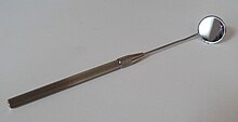
A laryngoscope (from the Greek larynx “ larynx ” and skopein “to look at”) or larynx mirror is a device
- for indirect viewing of the larynx ("indirect laryngoscopy"):
- a plane mirror with a handle ("larynx mirror"),
- a 70 ° to 90 ° magnifying endoscope
- a flexible fiber optic endoscope
- for direct viewing of the larynx ("direct laryngoscopy"):
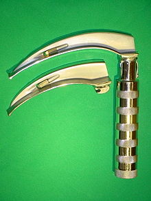
- a spatula-shaped ( intubation laryngoscope , intubation spatula ) instrument
- tubular ( surgical laryngoscope): device with lighting for direct viewing of the larynx.
The intubation laryngoscope is used as an aid for endotracheal intubation as part of anesthesia or ventilation therapy , the surgical laryngoscope is used in ear, nose and throat medicine to inspect and perform operations on the larynx.
When the patient is awake, the larynx can be viewed with the help of a plane mirror or an endoscope; this is practiced in laryngology ( ENT and phoniatrics ) and is known as indirect laryngoscopy . The direct laryngoscopy is only possible under general anesthesia or deep sedation or unconsciousness.
A intubating's daily work means the anesthesiologist in the operating room . It should also be included in every emergency kit , as it is required for intubation during resuscitation . Therefore, a laryngoscope with various blades should be available in every doctor's practice and in every rescue equipment manned by appropriately qualified personnel.
history
A laryngoscope-like instrument set was built in 1743 by the French obstetrician André Levret , who used it to visualize the vocal folds with a wide metal spatula.
After further attempts at laryngoscopy using the " light guide " by Philipp Bozzini (1806) or the "glottiscope" by Babington (London 1829) and by Robert Liston (1804), the Spanish opera singer Manuel García was the first to understand the anatomy of the larynx, especially its course of the muscles, studied with the help of a larynx mirror. In 1855 he managed to see his own larynx with a dentist's mirror and to observe the movements while singing. He has since been honored as the inventor of laryngoscopy. Garcia was primarily interested in laryngoscopy to clarify the functions of the larynx when singing, not so much in the medical aspect. Nevertheless, he received an honorary medical doctorate from the University of Königsberg .
Alfred Kirstein (1863–1922) demonstrated his first investigations on the internal structures of the larynx on April 23, 1895 . About three weeks later he presented his results to the Berlin Medical Society . Until now, laryngoscopy was only possible with a stalked mirror and reflecting light, i.e. as indirect laryngoscopy, as it was first used by Manuel Garcia, introduced into medicine by Ludwig Türck in 1858 and further developed by Johann Nepomuk Czermak . Kirstein's apparatus, on the other hand, was a combination of the Caspers electroscope developed by the Berlin urologist Leopold Casper (1859-1959) and an esophagoscope developed by Kirstein. Kirstein called the new device an autoscope and the examination method an autoscopy .
Larynx mirror
The larynx mirror is a 1.5 cm to 3 cm diameter mirror on a 15–20 cm long, thin handle angled by approx. 45 °, which is inserted into the patient's mouth up to the back of the throat. The organs examined ( pharynx and larynx ) are illuminated indirectly by a forehead mirror or headlamp of the examiner by deflecting the light beam via the larynx mirror.
Magnifying laryngoscope
For the technology s. endoscope
Magnifying laryngoscopes usually have a viewing angle of 70 ° or 90 °. The length of the endoscope tube is typically 15-20 cm.
Many magnifying laryngoscopes are equipped with a combined zoom and focusing device in order to be able to focus the image at different distances from the larynx or to select a different degree of magnification. For documentation of findings an image transfer using an endoscope camera (placed on the eyepiece) on video tape or digitally on hard disk, or more recently even be carried out with integrated into the endoscope camera chip ( video laryngoscopy ).
Areas of application
The magnifying laryngoscope is the main working device of the ENT doctor and phoniatrist working in laryngology . It will allow the investigation of the oropharynx , lower pharynx and larynx performed. In combination with stroboscopy or high-speed cameras , vibration analyzes of the vocal folds can be carried out.
Flexible (naso-pharyngo) laryngoscope
For the technology s. endoscope
Flexible endoscopes for use in this area have a working length of approx. 30–40 cm. The standard diameter is approx. 4 mm, for use in children and infants 1.5–2 mm. The lower endoscope section is usually equipped with a bending mechanism. The naso-pharyngo-laryngoscopes usually have no additional working channel.
Areas of application
In patients with gag reflex, it is not possible to examine the deeper parts of the throat and the larynx transorally ("via the mouth") with a larynx mirror or laryngoscope. In the case of children, this area is also not or not optimally visible due to a changed anatomy (e.g. higher larynx). The flexible endoscope is therefore used for the examination, which is inserted through the nose (ideally after local anesthesia ). For the flexible endoscopes, there is also the option of using endoscope cameras or endoscopes with camera chips on the endoscope tip.
Intubation laryngoscope
Structure and shapes
The intubation laryngoscope consists of a handle and a spatula. The spatulas are available in different sizes and shapes to suit the different anatomical conditions of the patient. The spatulas can be clipped onto the handle; they then protrude from the handle at a right angle . If the laryngoscope is not in use, the blade is attached to the handle or removed.
A light source is built into the spatula to improve visibility in the dark throat. The batteries required for this are usually built into the handle.
Warm and cold light
In the warm light version, there is a small light bulb in the spatula, which is supplied with power via a contact between the handle and the spatula. Always ensure that the pear is firmly seated, as this is a common problem that can lead to malfunction.
In the cold light version, the light source is integrated in the handle. The light reaches the spatula via a fiber optic cable .
Spatula shapes
macintosh
The Macintosh spatula (also Wisconsin-Foregger spatula) according to Robert Macintosh is slightly curved and, depending on the size, between 76 (size 0) and 176 mm (size 5) long. Sizes 3 and 4 are often used for adults.
The name is often misspelled as "McIntosh" - a mistake that runs through history: The name Macintosh was incorrectly spelled MacIntosh when the patent was applied for.
Miller
The Miller spatula is straight. The size corresponds roughly to that of a comparable Macintosh spatula. Due to its shape, it is preferred for children.
Dörges
The Dörges spatula is relatively straight, only the front part is curved. This shape of the spatula is only available in one size, but this spatula covers the majority of all patients. Weight markings (10 and 20 kg body weight) are attached to the spatula blade in order to provide children with a guide for the insertion depth. The aim of this type of spatula is to reduce the number of spatulas required in emergency cases to a minimum; in addition to the Dörges spatula, only one toddler spatula is required.
McCoy
The McCoy spatula is slightly curved, similar to the Macintosh spatula, but also offers the option of lifting the epiglottis with a movable, foldable tip.
Individual evidence
- ^ Anthony Jahn, Andrew Blitzer: A short history of laryngoscopie. (PDF; 484 kB). In: Log Phon Vocol. 21, 1996, pp. 181-185, online.
- ↑ C. Nemes: Evolution of anesthesia in Germany after 1846: The first fifty years to the beginning of local and regional anesthesia, endotracheal intubation and hyperbaric anesthesia. (PDF; 436 kB). Überlingen on Lake Constance online
- ^ Christian von Deuster: Larynx mirror. In: Werner E. Gerabek , Bernhard D. Haage, Gundolf Keil , Wolfgang Wegner (eds.): Enzyklopädie Medizingeschichte. de Gruyter, Berlin / New York 2005, ISBN 3-11-015714-4 , p. 730 f.
- ^ Joachim Gerlach: Carl and Dietrich Gerhardt. Contributions to the Würzburg medical history of the late 19th and early 20th centuries. In: Würzburg medical history reports. 4, 1986, pp. 105-134; here: p. 119.
- ^ M. Reinhard, E. Eberhardt: Alfred Kirstein (1863-1922) - pioneer of direct laryngoscopy Alfred Kirstein (1863-1922) - pioneer of direct laryngoscopy. In: Anästhesiol Intensivmed Emergency Med Schmerzther. 30 (4), 1995, pp. 240-246.


