Ventilation
The ventilator (or respiration ) is an artificial lung ventilation and is used to support or replacement of inadequate or no spontaneous breathing . Their life-sustaining function is a central component in anesthesiology , emergency medicine and intensive care medicine . Ventilation is provided in first aid as a donation of breath , in emergency medicine often with a resuscitator and in intensive care medicine with ventilators . In mechanical ventilation, a distinction is made between controlled ventilation and assisted ventilation . In addition, techniques of artificial ventilation are used in the conservative treatment of sleep apnea , for example CPAP therapy .
Clinical application
Ventilation, if necessary with additional oxygen supply, is used when spontaneous breathing fails ( apnea ) or becomes insufficient. This can occur during anesthesia , poisoning, cardiac arrest, neurological diseases or head injuries, as well as paralysis of the respiratory muscles due to spinal cord lesions or the effects of medication. A number of lung or chest injuries, as well as heart disease , shock, and sepsis may also require ventilation.
Depending on the clinical situation, ventilation can be continued for a few minutes, but also for months. While returning to spontaneous breathing under routine anesthesia is rarely a problem, weaning an ICU patient after prolonged ventilation is a difficult process that can take days or weeks.
Some patients with severe brain damage, spinal cord injuries or neurological diseases do not regain the ability to breathe spontaneously and therefore require constant ventilation ( home ventilation ).
The ventilation efficiency is monitored by observing the patient as well as by pulse oximetry , blood gas analysis and capnometry .
techniques
Positive and negative pressure ventilation
While the exchange of oxygen and carbon dioxide between the blood and the alveoli takes place by diffusion and does not require any external effort, the respiratory air must be actively supplied to the gas exchange through the airways . With spontaneous breathing, a negative pressure is created in the pleural cavity by the respiratory muscles. The resulting pressure difference between atmospheric pressure and intrathoracic pressure creates an air flow.
- This mechanism is imitated in (historical) negative pressure ventilation with iron lungs . The iron lung creates a negative pressure in a chamber that encloses the body and is sealed at the neck. Nowadays, only the cuirass ventilation is used to a certain extent in home ventilation, where a kind of vest creates a negative pressure in the chest.
- All modern ventilation techniques are positive pressure ventilation : Air is pressed intermittently into the lungs by external positive pressure and passively exhaled again after reaching the desired tidal volume or pressure.
As early as 1955, Jürgen Stoffregen was able to show that the same transpulmonary pressure difference exists in both procedures.
Mouth-to-mouth and bag ventilation
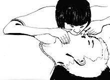
The simplest form of respiration is the respiration , which in layman resuscitation is applied. This means ventilation with the exhaled air of the helper, i.e. either "mouth-to-mouth" or "mouth-to-nose ventilation". This technique is limited, however, as it cannot provide oxygen-enriched air: Only 16 percent oxygen content can be achieved in this way; In comparison, room air has 21 percent oxygen, ventilators can achieve up to 100 percent oxygen. With mouth-to-mouth resuscitation, there is always a low risk of disease transmission due to direct contact with body fluids; this can be minimized by using a respiratory aid.
Professional helpers should therefore use aids such as the resuscitator , provided the technology is mastered. A resuscitator consists of a face mask that is placed over the patient's mouth and nose in order to achieve a tight seal; an elastic, compressible bag and a valve that directs the air flow. An oxygen source can be connected to a reservoir on the bag to achieve a higher concentration of oxygen. This simple technique can be sufficient to ventilate a patient with insufficient breathing or apnea for hours.
Mechanical ventilation using mechanical fans
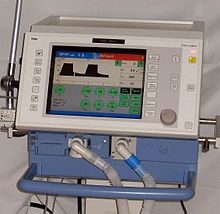
Ventilators are routinely used in anesthesiology and intensive care medicine . These fans enable a variety of different ventilation modes, ranging from assisted spontaneous breathing (ASB) to fully controlled ventilation. Modern fans allow continuous adaptation of the invasiveness according to the patient's condition.
In ventilated patients there is a tendency for alveoli to collapse ( atelectasis formation ). By using PEEP (positive end-expiratory pressure) one tries to keep the alveoli open at the end of a breathing cycle. PEEP is also used for diseases such as pneumonia , ARDS and pulmonary edema .
High-frequency oscillation ventilation ( HFOV ) is a special form of intensive care ventilation . A continuous flow system generates a continuous inflation pressure that enables the alveoli to be kept open. This is why this procedure is used in particular for hypoxemic lung failure ( ARDS ).
The iron lung no longer plays a role in modern medicine.
Non-invasive ventilation (NIV)
As an alternative to intubation (and thus the "invasive" ventilation) one can under certain conditions, non-invasive ventilation (English non-invasive ventilation , NIV ) are used. This is understood to mean mostly automatic (machine) ventilation using face masks, mouth-nose masks or ventilation helmets, in which no artificial airways such as endotracheal tubes or tracheal cannulas are inserted into the body as tubes and in which sedation is largely triggered (if the significant additional psychological Exposure, especially at the beginning of NIV) can be dispensed with, whereby disadvantages of invasive ventilation (e.g. infections in immunosuppressed patients) should be avoided.
Non-invasive forms of ventilation require an air-tight connection between the ventilator and airways, so that different face masks, mouth / nose masks, nasal masks (with the mouth closed) or full-head helmets are used depending on the patient. In the intensive care unit too, ventilator settings are used that can support existing spontaneous breathing (usually pressure-supported ventilation such as proportional assist ventilation ).
application areas
Non-invasive ventilation is used in acute respiratory distress due to the worsening of existing COPD , in pulmonary edema associated with increased carbon dioxide in the blood (although it can stabilize the hemodynamics in cardiogenic pulmonary edema with acute hypoxemic respiratory insufficiency ), pneumonia , asthma and in the treatment of respiratory insufficiency (primarily to reduce the work of breathing when the respiratory muscles fail and in the case of acute hypoxemic respiratory insufficiency after major surgical interventions, but also to reopen and stabilize atelectatic lung areas in hypoxic or hypoxemic lung failure ) as part of an intensive treatment, whereby in the case of pronounced atelectasis formation (and intrapulmonary right-left shunt ) the effectiveness is limited. By using non-invasive ventilation in good time (as a therapy attempt - over one to two hours - always justified if there are no contraindications), the insertion of a ventilation tube into the windpipe can be avoided. NIV is therefore also suitable for weaning from invasive ventilation and for avoiding re-intubation.
In addition, there are also applications for non-invasive ventilation in the field of home ventilation . The CPAP therapy z. B. in sleep apnea syndrome can be counted in the broadest sense of the NIV, whereby technically the CPAP represents only a passive maintenance of an overpressure in the exhalation phase. Ventilation in the sense of active support for inhalation is not CPAP.
For the out-of-hospital emergency treatment of seriously injured people with insufficient oxygen saturation of the blood, invasive ventilation under anesthesia is recommended, as this ensures better protection of the patient from pain and stress.
Contraindications
The NIV must not be used in the absence of spontaneous breathing, obstruction of the airways, bleeding in the gastrointestinal tract, intestinal obstruction and in a coma not caused by hypercapnia.
Advantages and disadvantages
As can be seen from the table, the advantage of NIV is that no tubes have to be inserted into the airways, thereby reducing complications such as pneumonia in particular. The main disadvantage of NIV is the lack of protection of the lungs from gastric juice aspiration, which is why NIV should not be used in patients at risk of aspiration.
| Complications / special features | invasive ventilation (e.g. intubation) | non-invasive ventilation (NIV) |
|---|---|---|
| ventilation pneumonia | Increase in risk from the 3rd to 4th day of ventilation | Rare |
| additional breathing work due to the hose | yes (during spontaneous breathing and in the case of insufficient tube compensation) | No |
| Damage to the windpipe or larynx | Yes | No |
| Sedative drug | often necessary | rarely necessary |
| temporary pause | rarely possible | often possible |
| to eat and drink | hardly possible (only with tracheostomy) | Yes |
| Speak | No | Yes |
| Sit | rarely possible | Yes |
| Weaning problems | 10-20% | no |
| Access to the airways | directly | difficult |
| Pressure points on the face | No | occasionally |
| CO 2 rebreathing | No | Rare |
| Escape of ventilation air | barely | mostly available |
| Swallowing air | barely | occasionally |
Table adapted according to
Technology and application
In the case of acute worsening of COPD or asthma, the respiratory muscles are usually overwhelmed by excessive resistance to inhalation. NIV should therefore actively support inhalation. Non-invasive ventilation can generally be performed with any modern ventilator. The pressure support during the inhalation phase should be set on the ventilator so that the total inhalation pressure in the airways is 15–25 (or the support pressure 5 to 8) cmH20. During the exhalation phase, a pressure between 3 and 9 cmH20 (PEEP) should be maintained. This helps the patient to inhale and compensates for the intrinsic PEEP caused by the disease.
In the event of an acute deterioration in respiratory function due to pulmonary edema or pneumonia, the alveoli are no longer ventilated and collapse. Therefore, with NIV, a pressure of between 10 and 15 cm H20 should be maintained in the exhalation phase (PEEP) in order to open or keep the alveoli open. Pressure support during inhalation is only necessary if there is secondary exhaustion of the respiratory muscles.
Various ventilators with NIV functions are available for intensive care units and specialized nursing homes. For the use of NIV outside the hospital in the rescue service, only a few special ventilators are available so far. B. Dräger Oxylog 2000 + / 3000, Weinmann Medumat Transport, Cardinal Health LTV 1200.
High-flow systems, nasal high-flow oxygen therapy (NHF)
As a pure CPAP system without the possibility of pressure support, there are high-flow systems that generate a PEEP via a high gas flow (e.g. Vygon CPAP Boussignac valve). Similarly, if NIV cannot be tolerated or if it cannot be performed adequately due to anatomical features before switching to invasive ventilation, the use of nasal (applied through a nasal cannula) high-flow oxygen therapy (NHF) can be used at 40 to 60 liters of oxygen / minute (instead of the conventional 2 to 12 l / min) should be attempted (if the patient improves, a reduction to a maximum of 20 l / min is recommended). The NHF thus represents both an alternative to NIV and to conventional oxygen therapy. Clinical study results are available, especially for the use of NHF oxygen therapy in hypoxic lung failure after surgical interventions, and therapy attempts with this method are recommended (according to the 2015 guideline attached mild to moderate lung failure as well as an alternative to NIV in acute hypoxemic respiratory failure in patients after cardiac surgery and in immunosuppressed patients). Effective mechanisms are discussed: the increased oxygen concentration in the inhaled air, a leaching of carbon dioxide from the anatomical dead space, the reopening (recruitment) of atelectatic lung areas through continuous airway pressure as well as the heating and humidification of the administered breathing gas.
Securing the airway
Mechanical ventilation can only be successful and safe if the patient's airways are kept open (secured) and if air can flow freely into and out of the lungs. In addition, leakages must be avoided (or compensated for by a higher breathing gas flow) so that the air flow and pressure conditions correspond to the set values.
Another risk is aspiration , in which stomach contents enter the lungs via the esophagus ( esophagus ) and windpipe ( trachea ). Obstruction of the airways or the acidity of the stomach contents can lead to severe impairment of lung function up to ARDS .
Measures to secure the airway depend on the situation of the individual patient, but the most effective protection is provided by endotracheal intubation . Alternatives are supraglottic airway aids that are located above the glottis . Available are laryngeal mask , laryngeal tube and Combitubus , they are often used in difficult or not possible intubation as an alternative. Non-invasive ventilation is provided through a mask or a special helmet.
The tracheotomy (incision of the windpipe) is a surgical procedure in which the soft tissue of the neck provides access to the windpipe . Indications for a tracheotomy can be the need for long-term ventilation, neurological diseases with disorders of the swallowing reflex, radiation treatment of the head or neck, or laryngeal paralysis.
Ventilation-induced lung damage and lung-protective ventilation
In general, the ventilator's prognosis is determined by the underlying disease and its response to therapy. But ventilation itself can also cause serious problems, which in turn extend the stay in an intensive care unit and can sometimes lead to permanent damage or even death. Ventilation technology is therefore geared towards preventing this type of ventilation damage. This includes keeping the ventilation time as short as possible.
Infectious complications, especially pneumonia , occur more frequently in patients who remain ventilated for more than a few days. Endotracheal intubation undermines the natural defense mechanisms against lung infections, in particular the process of "mucocillary clearance ". This continuous transport of secretions from the lungs into the upper airways serves to remove bacteria and foreign bodies. The deactivation of this mechanism due to intubation is considered to be the main factor in the development of pneumonia.
There are indications that oxygen in higher concentrations (> 40%) can in the long run even damage the lung tissue of ventilated patients. It is therefore advisable to set the lowest appropriate oxygen concentration. However, in patients with severe pulmonary gas exchange disorders, a high oxygen concentration may be necessary for survival.
Most ventilation modes rely on the application of positive pressure to the lungs. The tissue of diseased lungs can be additionally damaged by the resulting mechanical stress (overstretching, shear forces, too high peak pressures, too low PEEP, too high ventilation volumes) as well as by inflammatory processes. The resulting deterioration in pulmonary gas exchange can then in turn require even more aggressive ventilation.
"Lung-protective ventilation" is a collective term for strategies to minimize ventilation-induced lung damage. Many of them are based on ventilator settings to avoid overextension and cyclical collapse of the lungs.
Basic principles of mechanical ventilation
A fundamental distinction is made between controlled (mandatory) ventilation (CMV), in which the patient's work of breathing is completely taken over, and supported (augmented) spontaneous breathing, with the respiratory rate and depth of breath, i.e. the tidal volume (V T ), by the Patients are controlled.
Mandatory ventilation (MV) can be divided into volume-controlled mechanical ventilation , pressure-controlled mechanical ventilation and demand mechanical ventilation , which differ in terms of different inspiratory and expiratory controls:
- Volume control defines how much air the patient inhales and the delivery of this preselected volume ends inspiration. This results in pressure conditions in the lungs , which result from their condition and the inhaled volume . Forms of ventilation are, for example, CMV ventilation (controlled ventilation) and (S) IMV .
- Pressure control: The pressure-controlled ventilation determines how much pressure may prevail in the lungs and subordinates the tidal volume. The inspiration ends here when the preselected pressure is reached. That is, the maximum pressure in the lungs is constant while the volume varies. This form can also be defined with CMV and SIMV .
- Demand ventilation is a hybrid of the two above; both the volume to be inhaled and a pressure limit can be set. Volume inconsistencies exist in this form; Self-ventilation of the patient is possible but not mandatory. BiPAP / BIPAP has established itself as the preferred form of ventilation . Depending on the manufacturer of the ventilator, BiPAP is also known as Bi-Vent , BiLevel or BIPHASE .
- Flow control: If the inspiration flow falls below a specified level, inspiration ends.
- Time control: After a preselected time has elapsed, inspiration or expiration is ended.
- Patient trigger: After detection of a spontaneous attempt to inhale by the patient, expiration is ended.
Augmented spontaneous breathing can be found in CPAP , pressure support, and proportional pressure support .
- CPAP does not provide breathing assistance. The patient has to breathe independently, he is only provided with a pressure in the ventilation system that he can use. However, the constant positive airway pressure can increase the gas exchange surface in the lungs of the spontaneously breathing patient.
- Pressure support provides assistance with breathing. This assistance is constant, i.e. it is present to the same extent with every breath. ASB is the method of choice. The proportional pressure support (PAV) is an adapted breathing assistance, the support depends on the patient and is inconsistent, i.e. different with each breath.
Intermittent mandatory ventilation is a hybrid of mandatory and augmented ventilation. The ventilated person controls the frequency and depth of breath. As a rule, breathing support is provided by ASB.
Nomenclature of mechanical ventilation and respiratory support
Due to a lack of standardization in the field of mechanical ventilation and the large number of providers, various names for forms of ventilation have emerged. Some of these terms refer to identical forms of ventilation. However, they can also have different characteristics and meanings from manufacturer to manufacturer. The following list is not exhaustive:
- APRV Airway Pressure Release Ventilation
- ASB Assisted Spontaneous Breathing - assisted spontaneous breathing
-
ASV
- Adaptive Support Ventilation - closed-loop ventilation, a further development of MMV
- Adaptive servo ventilation a non-invasive ventilation for central sleep apnea syndrome
- ATC Automatic Tube Compensation - Automatic tube compensation
- BiPAP Biphasic Positive Airway Pressure - two-phase positive breath pressure support
- BiPAP Bilevel Positive Airway Pressure - two-phase positive breath pressure support in NIV
- CMV Continuous Mandatory Ventilation - continuous, fully mechanical ventilation (also Controlled Mandatory Ventilation - controlled ventilation)
- CPAP Continuous Positive Airway Pressure - continuous positive airway pressure
- CPPV Continuous Positive Pressure Ventilation - continuous positive pressure ventilation
- EPAP Expiratory Positive Airway Pressure - positive expiratory airway pressure
- HFPPV High Frequency Positive Pressure Ventilation - high frequency positive pressure ventilation
- HFOV High Frequency Oscillatory Ventilation - high frequency ventilation
- HFV High Frequency Ventilation - high frequency ventilation
- ILV Independent Lung Ventilation - side-separated positive pressure ventilation
- IMV Intermittent Mandatory Ventilation - intermittent total substitution of individual breaths
- IPAP (absolute) Inspiratory Positive Airway Pressure - positive inspiratory airway pressure
- IPAP (relative) Inspiratory Pressure Above PEEP - positive inspiratory ventilation pressure above the PEEP level
- IPPV Intermittent Positive Pressure Ventilation - intermittent positive pressure ventilation
- IPV Intrapulmonary Percussive Ventilation - high-frequency open positive pressure ventilation, invasive, non-invasive (synonym: HFPV-High Frequency Percussive Ventilation)
- IRV Inversed Ratio Ventilation - ventilation with reversed breathing phases / with reversed time ratio
- LFPPV Low Frequency Positive Pressure Ventilation - low frequency positive pressure ventilation
- MMV Mandatory Minute Volume - (default) machine minute volume
- NIV non-invasive ventilation (non-invasive ventilation)
- NPPV Noninvasive Positive Pressure Ventilation: - Noninvasive positive pressure ventilation
- NPV Negative Pressure Ventilation (negative pressure ventilation, negative pressure ventilation ); z. B. in the cuirass method
- PAV Proportional Assist Ventilation - proportional pressure-assisted ventilation
- PC Pressure Control - pressure controlled, fully mechanical ventilation
- PCMV (P-CMV) Pressure Controlled Mandatory Ventilation - pressure controlled, fully mechanical ventilation
- PLBV - Pursed Lip Breathing Ventilation
- (A) PCV (Assisted) Pressure Controlled Ventilation - pressure controlled, fully mechanical ventilation
- PEEP Positive End Expiratory Pressure - positive end expiratory pressure
- PNPV Positive Negative Pressure Ventilation - alternating pressure ventilation
- PPS Proportional Pressure Support - proportional pressure-supported ventilation (Draeger), see also PAV
- PRVC Pressure Regulated Volume Controlled; - Pressure-regulated volume-controlled ventilation (Weinmann), see Draeger = autoflow ventilation
- PSV ventilation Pressure Support Ventilation - Pressure-supported spontaneous breathing, see also ASB
- S-CPPV Synchronized Continuous Positive Pressure Ventilation - synchronized continuous positive pressure ventilation
- S-IPPV Synchronized Intermittent Positive Pressure Ventilation - synchronized intermittent positive pressure ventilation
- (S) IMV (Synchronized) Intermittent Mandatory Ventilation - (synchronized) intermittent mechanical ventilation
- VCMV (V-CMV) Volume Controlled Mandatory Ventilation - volume-controlled, fully mechanical ventilation
- VCV Volume Controlled Ventilation - volume controlled, fully mechanical ventilation
- ZAP Zero Airway Pressure - spontaneous breathing under atmospheric pressure
Ventilation parameters
The setting of the ventilation parameters are based on size, weight, and clinical condition of the patient and will be based on clinical , vital signs , blood gases , pulse oximetry and capnometry validated.
The most important ventilation parameters to be monitored by means of monitoring include:
Oxygen concentration
The oxygen concentration can (depending on the manufacturer) be set within the limits of 21% to 100% of the gas mixture. It is given both in percent and as FiO 2 , the inspiratory oxygen fraction (decimal value). Ventilation with 100% oxygen (FiO 2 = 1.0) is carried out, for example, in life-threatening conditions, during preoxygenation before intubation or before endotracheal suctioning of the patient.
Respiratory rate
The breathing rate describes the number of ventilation cycles applied per minute; the usual settings are between 8 and 30 / min. Devices specially developed for neonatology achieve much higher ventilation frequencies. The setting range here is usually 8 to 150 rpm.
The respiratory rate can be set to an absolute value or to a minimum value. The setting to a minimum value is used to perform assisted ventilation.
Tidal volume
The tidal volume (VT) corresponds to the air volume per breath, the tidal volume (AZV), and is the set volume that is to be applied per breath. The tidal volume of independent spontaneous breathing is around 0.5 l in adults. With volume-controlled ventilation (VCV), this value can be set precisely to suit the patient, using the rule of thumb of 7–8 ml per kilogram of ideal body weight. This parameter is the most important parameter in volume-dependent ventilation.
Minute ventilation
The respiratory minute volume indicates the volume that is administered per minute. It depends to a large extent on the type of ventilation chosen and must be adapted to the needs of the patient.
- ,
where is the respiratory rate.
Inspiratory flow (flow)
The flow is the value for the amount of gas flowing into the patient in relation to time. A high flow ensures rapid ventilation, a lower flow ensures better distribution of the breathing gases in the lungs. The inspiratory flow can be constant, decreasing (decelerating) or increasing (accelerating). The advantages of these different flow forms have been discussed very controversially for 40 years.
Maximum inspiratory pressure
Volume-controlled ventilation with constant flow results in a short-term peak pressure which drops to what is known as the plateau pressure in the plateau phase (inspiratory hold). With volume-controlled ventilation, the inspiratory ventilation pressures are a degree of freedom and depend on the tidal volume, resistance and compliance of the lungs.
The set pressure (P max) is quickly reached with pressure-controlled ventilation through a high flow at the beginning of the inspiration phase, then the flow decreases again. The ventilation gas mixture can then be supplied by the device until the inspiration time has expired. Here the tidal volume is the degree of freedom. In this form, the flow is referred to as a decelerating (slowing down) flow.
Adjuvant measures and therapies (examples)
- Positioning drainage
- Endotracheal or bronchoscopic suction
- Physiotherapy ( respiratory gymnastics or respiratory physiotherapy ; see Pneumological Rehabilitation # Individual Measures )
- Prone position
- Breathing gas conditioning
- Mini tracheotomy
- Extracorporeal membrane oxygenation
- pECLA - Pumpless extracorporeal membrane oxygenation
- Liquid ventilation (Engl. Liquid ventilation )
- Side-separated ventilation ( independent lung ventilation ); see Endotracheal Intubation
history
Early descriptions of various ventilation measures can be found in Hippocrates , Avicenna and Paracelsus . From the 1st century BC Doctors working in Rome ( Asklepiades of Bithynia ) even report a tracheotomy . In 1763 Smellie used a flexible metal tube for intubation of the trachea, Fothergill used a bellows for help. The first iron lung was built in 1876 and was to be of great importance well into the 20th century. Laryngoscopy emerged around 1900 and paved the way for the endotracheal intubation that is common today. The Pulmotor has been sold and used since 1908 . The back pressure arm pull method was used until around the middle of the 20th century . Around this time the first mechanical respirators from Puritan Bennett, Bird, Blease, Dräger , Engström, Emerson etc. were developed. From the end of the 1980s, devices were developed that also met the demands of modern ventilation for newborns and even premature babies.
See also
literature
- F. Bremer: 1 × 1 of ventilation. 4th edition. Lehmanns Media, Berlin 2014, ISBN 978-3-86541-577-6 .
- Harald Keifert: The ventilation book. Invasive ventilation in theory and practice. 4th edition. WK reference books, Elchingen 2007, ISBN 978-3-9811420-0-6 .
- Walied Abdulla: Interdisciplinary Intensive Care Medicine. Urban & Fischer, Munich a. a. 1999, ISBN 3-437-41410-0 , pp. 5-12.
- R. Larsen, Thomas Ziegenfuß : Ventilation. Basics and practice. 2nd Edition. Springer, Heidelberg 1999, ISBN 3-540-65436-4 .
- Wolfgang Oczenski, Alois Werba, Harald Andel: Breathing - breathing aids. Respiratory physiology and ventilation technology. 6th edition. Thieme, Stuttgart 2003, ISBN 3-13-137696-1 .
- Eckhard Müller: Ventilation. Scientific basics, current concepts, perspectives Thieme, Stuttgart 2000, ISBN 3-13-110241-1 .
- H. Becker, B. Schönhofer, H. Burchardi: Non-invasive ventilation. Thieme, Stuttgart 2002, ISBN 3-13-137851-4 .
- S. Schäfer, F. Kirsch, G. Scheuermann, R. Wagner: Specialist care ventilation. Elsevier, Munich 2005, ISBN 3-437-25182-1 .
- Martin Bachmann: Ventilation. Jörg Braun, Roland Preuss (Ed.): Clinical Guide Intensive Care Medicine. 9th edition. Elsevier, Munich 2016, ISBN 978-3-437-23763-8 , pp. 95-130.
- Ernst Bahns: It all started with the Pulmotor. The history of mechanical ventilation. Drägerwerk, Lübeck 2014.
Web links
- Information at emedicine.com
- A simulator that enables controlled forms of ventilation to be trained on a respirator.
Individual evidence
- ↑ Non-invasive ventilation as a therapy for acute respiratory failure. Edited by the German Society for Pneumology and Respiratory Medicine. In: AWMF online. 2015.
- ↑ AWMF: S3 guideline on invasive ventilation and the use of extracorporeal procedures in acute respiratory insufficiency. (December) 2017, Chapter 2 ( Indications for invasive ventilation ).
- ↑ a b c d e f g h i Rolf Dembinski: Non-invasive forms of ventilation . In: Anaesthesiology & Intensive Care Medicine . tape 60 , no. 6 . Aktiv Druck & Verlag, June 2019, p. 308–315 , doi : 10.19224 / ai2019.308 ( ai-online.info [PDF; 189 kB ; accessed on April 4, 2020]).
- ↑ a b c S3 guidelines: Non-invasive ventilation as a therapy for acute respiratory insufficiency as of 2008 ( awmf.org ).
- ↑ Bronchial asthma and chronic obstructive pulmonary disease with acute exacerbation. In: The anesthesiologist . Volume 58, No. 6, June 2009, pp. 611-622, doi: 10.1007 / s00101-009-1536-x .
- ↑ P. Kruska, T. Kerner: Acute respiratory insufficiency - Preclinical therapy of obstructive ventilation disorders. In: anesthesiology, intensive care medicine, emergency medicine, pain therapy . 46, 2011, pp. 726-733, doi: 10.1055 / s-0031-1297179 .
- ↑ O. Roca et al. a .: Current evidence for the effectiveness of heated and humidified high flow nasal cannula supportive therapy in adult patients with respiratory failure. In: Critical Care. Volume 20, 2016, Item No. 109, doi: 10.1186 / s13054-016-1263-z .
- ↑ Ernst Bahns: It all started with the Pulmotor. The history of mechanical ventilation. Drägerwerk, Lübeck 2014, p. 66 f. ( New ventilation technology with EV-A ).
- ↑ M. Baum: Technical basics of ventilation. In: J. Kilian, H. Benzer, FW Ahnefeld (ed.): Basic principles of ventilation. Springer, Berlin a. a. 1991, ISBN 3-540-53078-9 , 2nd, unchanged edition, ibid 1994, ISBN 3-540-57904-4 , pp. 185-200; here: pp. 189–198.
- ↑ D. Weismann: Forms of ventilation. In: J. Kilian, H. Benzer, FW Ahnefeld (ed.): Basic principles of ventilation. Springer, Berlin a. a. 1991, ISBN 3-540-53078-9 , 2nd, unchanged edition, ibid 1994, ISBN 3-540-57904-4 , pp. 201-211, here: pp. 203-205.
- ↑ The influence of controlled mandatory ventilation (CMV)… PMID 9466092
- ↑ D. Weismann: Forms of ventilation. In: J. Kilian, H. Benzer, FW Ahnefeld (ed.): Basic principles of ventilation. Springer, Berlin a. a. 1991, ISBN 3-540-53078-9 ; 2nd, unchanged edition, ibid. 1994, ISBN 3-540-57904-4 , pp. 201-211, here: pp. 203-208.
- ↑ W. Koller, TH Luger, Ch. Putensen, G. Putz: Blood purifying procedures in intensive care medicine. In: J. Kilian, H. Benzer, FW Ahnefeld (ed.): Basic principles of ventilation. Springer, Berlin a. a. 1991, ISBN 3-540-53078-9 , 2nd, unchanged edition, ibid 1994, ISBN 3-540-57904-4 , pp. 404-419, here: pp. 404-407.
- ↑ Vigaro. In: novamed.de. Retrieved October 24, 2017 .
- ↑ D. Weismann: Forms of ventilation. In: J. Kilian, H. Benzer, FW Ahnefeld (ed.): Basic principles of ventilation. 2nd, unchanged edition. Springer, Berlin a. a. 1994, ISBN 3-540-57904-4 , pp. 201-211, here: pp. 209 f. ( Ventilation monitoring ).
- ^ Inspiratory oxygen fraction.
- ↑ Ernst Bahns: It all started with the Pulmotor. The history of mechanical ventilation. Drägerwerk, Lübeck 2014, pp. 48–51.


