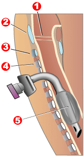
Tracheotomy

(2) cricothyroid ligament
(3) the cricoid cartilage
(4) trachea
(A) localization of the Koniotomie
(B) Localization of the tracheotomy
The tracheotomy (from Greek τραχεῑα from τραχύς trachýs , 'rough', 'hard', and τομή tomē , 'cut') - also tracheotomy - is a surgical procedure that has been practiced since ancient times, in which the soft parts of the neck provide access to the windpipe (tracheostoma ) is created. Indications for a tracheotomy can be, for example, the need for long-term ventilation after accidents or operations, neurological diseases with disorders of the swallowing reflex, radiation treatment of the head or neck or laryngeal paralysis. Even patients who have had their larynx removed completely ( laryngectomy ) have a tracheostoma.
Colloquially, a tracheal incision is also incorrectly understood to be a life-saving measure in emergency medicine, the cricothyrotomy , although the actual windpipe is not affected (a so-called emergency tracheotomy is replaced by the faster and safer measures of endotracheal intubation or coniotomy). This is done as a last resort in the event of an acutely life-threatening complete obstruction or an occlusion of the upper airway (due to occlusion or obstruction ) to prevent the patient from suffocating.
history
A tracheotomy is shown in images of Ancient Egypt .
Asklepiades of Bithynia (around 95 BC) is considered the inventor of the tracheotomy known by name . Avicenna (died 1037) recommends tracheotomy in the event of life-threatening obstructions of the airways by foreign bodies . The first concrete reports on performing a tracheotomy can be found in a work by the Italian surgeon and anatomist Giulio Casseri (Julius Casserius, 1552–1616) Pierre Fidèle Bretonneau (1821) is also named as the inventor of the tracheotomy . His French compatriot Armand Trousseau also performed tracheotomies.
Surgical methods
Percutaneous puncture and dilatation tracheotomy
The trachea is punctured from the outside with a hollow needle and a guide wire is advanced. The correct position of the wire in the windpipe is checked endoscopically and the access is then widened over the guide wire with plastic dilators until a breathing cannula fits into it. This simple procedure is often used in intensive care units on ventilated patients when artificial ventilation is expected over a long period of time, but the prospect that a breathing cannula will not have to be worn continuously.
Surgical tracheotomy
Classic (plastic) tracheotomy
With this method, a clear surgical access to the trachea is created in the operating room, often parts of the thyroid gland are cut and blood vessels are tied off. Then the windpipe is opened and the tracheal tube is inserted from the outside through the soft tissue of the neck.
The resulting tracheostoma is larger and more stable than with percutaneous puncture tracheotomy and also allows the tracheostomy tube to be changed routinely. However, the tracheostoma is not permanently stable: If no tube is used for a long time, it shrinks and usually closes. Changing the cannula is sometimes no longer possible just a few minutes after removing the cannula. If it is to be expected that an artificial respiratory opening will have to be worn for a long time, a "plastic tracheostoma" is created. A part of the trachea is opened like a window sash and sutured to the skin of the neck. The result is a stable breathing channel without a wound surface. The patient can safely change the cannula himself. Such a plastic tracheostoma usually has to be closed by a new operation when it is no longer needed.

Tracheostomy for laryngectomy
After a complete removal of the larynx ( laryngectomy ), the trachea, separated from the lower part of the larynx, is permanently relocated to the outside and sewn into the skin of the neck. Medically correct, this procedure is called a tracheostomy. Since the larynx is removed, the procedure cannot be reversed. Laryngectomized people no longer have vocal cords and have to learn a substitute voice in the course of speech therapy treatment. This can be achieved through aids such as an electronic speaking aid or a so-called voice prosthesis . The latter is usually used as standard during the larynx operation between the trachea and esophagus .
Advantages of tracheotomy over intubation
- With long-term ventilation, the risk of damage to the vocal cords and windpipe is minimized.
- By switching off the upper airways and the resulting reduction in the " dead breathing space ", breathing becomes easier for the patient and thus weaning from the ventilator is made easier or even possible in the first place.
- Oral care and hygiene are made much easier.
- The patient no longer has the feeling of permanently having a foreign body in their mouth.
- Compared to intubation, the patient requires considerably less or no analgesic sedation.
- Oral feeding is possible.
Disadvantages of the tracheotomy compared to normal breathing through the mouth and nose
After a tracheotomy, the air no longer flows through the upper airways, but directly into the windpipe and lungs .
- The air we breathe is no longer moistened in the nose and no longer reaches the olfactory nerves, i.e. H. Tracheotomized people can no longer smell and therefore only taste to a limited extent.
- The upper airway cleaning function is switched off.
- Increased secretion formation due to irritation of the trachea (foreign body stimulus from the cannula).
All of the disadvantages mentioned here naturally also apply to normal orotracheal intubation. So you cannot help balance tracheotomy versus intubation.
To counter some of these disadvantages, "artificial noses" are used. This is an HME (abbreviation for: "Heat and Moisture Exchanger"). This is a plastic filter that contains water-binding material. The filter is placed on the tube or tracheostomy tube. It ensures that a large part of the existing air humidity remains in the airways and the inhaled air is humidified and warmed.
Breathing cannulas
After a tracheotomy, breathing cannulas (tracheostomy tubes) are inserted into the patient to keep the tracheostoma open and, if necessary, to ventilate through an inflatable "block" or "cuff" and to prevent pharyngeal secretions from reaching the lungs. Special forms of breathing cannulas ("speaking cannulas") also allow voices to be formed through openings in the cannula tube and speaking valves. Here, air flows through the larynx when you exhale. Cannulas are made of plastic (polyvinyl chloride, PVC) or metal (silver or nickel silver ). The advantage of metal cannulas is that they have a larger inner diameter with the same outer diameter and are less clogged with secretion. Inner cannulas ("soul") allow cleaning without having to change the entire cannula. The picture shows a size 8 PVC cannula (88 mm length and 11 mm outer diameter) with a block sleeve and inner cannula. The guide rod (obturator) is used for easier insertion.
Tracheostomy tube weaning
Breathing through a tracheostomy tube bypasses the functions of the nose . Therefore it should be limited to the necessary time. A decannulation plug can be used to wean a tracheostomized patient from the tracheostomy tube. If the patient's larynx is still intact, breathing through the tracheostoma is blocked for a certain period of time, under medical observation. The patient is required to breathe naturally through the mouth or nose for a certain period of time. Natural breathing can initially be strenuous for the person concerned, so the tracheotomist must gradually get used to natural breathing.
Closure of the tracheostoma
If the tracheostomy is no longer required, it must be closed. After a surgical tracheotomy, this is usually done surgically. In preparation, the opening is masked for about six weeks. If the channel is not closed after this time, the closure is carried out surgically. The trachea is sutured and the outer skin is closed. Possible complications are emphysema , wound infection, and bleeding . The history of the area of surgery, such as tissue damage from radiation, should be considered.
literature
- Christian Beyer, Thomas Kerz: From tracheotomy to decannulation: a transdisciplinary manual . Lehmanns Media, Berlin 2013, ISBN 978-3-86541-512-7 .
- Biesalski, Fahl: Tracheal cannula Instructions for use Ref. 832/03 February 2014.
Remarks
- ↑ The word formation is derived from the feminine τραχεῖα tracheia .
- ↑ Provision in case of swelling
- ^ Teacher of Adriaan van de Spiegel
- ↑ With elastic tape and a pressure plate to relieve pressure when speaking
Web links
- New tracheostomy techniques in the intensive care unit. (PDF)
- Tracheostomy Nursing Guide. NOAH Foundation
- tracheotomie-online.de - further information on the procedure, history and other aspects of the tracheotomy
- Tracheotomy yesterday and today . International Symposium 2006 at the University of Greifswald
- Tracheotomy . Doccheck Flexikon
Individual evidence
- ^ Wilhelm Gemoll : Greek-German school and hand dictionary . G. Freytag Verlag / Hölder-Pichler-Tempsky, Munich / Vienna 1965.
- ^ Pschyrembel . Medical dictionary. 257th edition. Walter de Gruyter, Berlin 1993, ISBN 3-933203-04-X , p. 1551 .
- ↑ Christoph Weißer: Tracheotomy. In: Werner E. Gerabek , Bernhard D. Haage, Gundolf Keil , Wolfgang Wegner (eds.): Enzyklopädie Medizingeschichte. De Gruyter, Berlin / New York 2005, ISBN 3-11-015714-4 , p. 869.
- ^ Rainer Fritz Lick , Heinrich Schläfer: Accident rescue. Medicine and technology . Schattauer, Stuttgart / New York 1973, ISBN 978-3-7945-0326-1 ; 2nd, revised and expanded edition, ibid 1985, ISBN 3-7945-0626-X , p. 150 f.
- ↑ Not for first aiders: cricothyrotomy and tracheotomy. Retrieved September 6, 2016 .
- ↑ Forgotten Knowledge: Did the Ancient Egyptians Know Intensive Care Medicine? . German pharmacist newspaper. Retrieved February 16, 2018.
- ↑ Ludwig Brandt, Michael Goerig: The history of the tracheotomy . Part 1. In: The anesthesiologist. Volume 35, 1986, pp. 279-283.
- ↑ Heinrich Schipperges (†): Ibn Sīnā (= abū ʿAlī al-Ḥusain ibn ʿAbd Allāh ibn Sīnā al-Qānūnī = Avicenna). In: Werner E. Gerabek , Bernhard D. Haage, Gundolf Keil , Wolfgang Wegner (eds.): Enzyklopädie Medizingeschichte. De Gruyter, Berlin / New York 2005, ISBN 3-11-015714-4 , pp. 1334–1336, here: p. 1335.
- ↑ Burkhard Kramp, Steffen Dommerich1: Tracheostomy cannulas and voice prosthesis . In: GMS Curr Top Otorhinolaryngol Head Neck Surg , March 10, 2011, doi: 10.3205 / cto000057 , PMC 3199818 (free full text)
- ↑ Karl Wurm, AM Walter: Infectious Diseases. In: Ludwig Heilmeyer (ed.): Textbook of internal medicine. Springer-Verlag, Berlin / Göttingen / Heidelberg 1955; 2nd edition, ibid. 1961, pp. 9-223, here: p. 87.
- ↑ Armand Trousseau: You Tubage de la glotte et de la tracheotomy. Paris 1851.
- ↑ Brief instructions for cleaning and caring for Rüsch Care tracheostomy tubes, p. 3 (PDF) Archived from the original on May 13, 2013. Info: The archive link was inserted automatically and has not yet been checked. Please check the original and archive link according to the instructions and then remove this notice. Retrieved April 28, 2017.
- ↑ Video Tracheotomy ( fr ) Retrieved January 8, 2017.
- ↑ LK Eberhardt: The dilated tracheotomy in the anesthesiological intensive care unit . urn : nbn: de: bsz: 289-vts-68212
- ↑ Artificial humidification . Retrieved January 8, 2017.
- ↑ S. Wilpsbäumer, L. Ullrich: Intensivpflege (= Thiemes care ). Georg Thieme, Stuttgart 2009, ISBN 978-3-13-500011-4 , p. 1440 .
- ^ Berthold Milius: Tracheostoma. Retrieved December 26, 2018 .
- ↑ a b Andreas Fahl: Instructions for use LARYVOX® SEAL. Fahl Medizintechnik, accessed on June 10, 2020 (German, English).
- ↑ a b Tracheostoma closure. Prolife, accessed June 14, 2020 .

