Thoracoscopy
The thoracoscopy refers to a surgical in the way chest cavity to see and the pleura ( pleura ) to assess and introduce with the same or more access instruments or drugs.
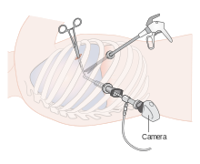
The instruments usually consist of a tube with a trocar that is inserted through the chest wall. Before this, a pneumothorax is created by opening the chest cavity . The lungs collapse due to the lack of negative pressure in the chest cavity. Since the lungs no longer occupy the entire chest cavity, the resulting free space in the chest cavity can be seen. Ventilation takes place with a double-lumen tube .
The procedure is part of minimally invasive surgery , which avoids opening the chest ( thoracotomy ). This results in significant advantages for patients, such as significantly reduced postoperative pain, fewer complications and a shorter hospital stay.
Video-assisted thoracoscopy

A further development is thoracoscopy with video support, video-assisted thoracoscopy ( English video-assisted thoracoscopic surgery , VATS ). Depending on the type of procedure, this can also be carried out with flexible endoscopes .
Up until the 1990s, these interventions were often only used for diagnosis or were used to treat diseases such as pneumothorax , pleural effusion or pleural empyema . Thanks to the development of reliable methods for closing the lung tissue and blood vessels, minimally invasive thoracic surgery can now also be used for the removal of a disease-bearing unit of the lung ( lung resection ) or the removal of lymph node metastases for histological examination. Most often, the operation is used to confirm or exclude a cancer diagnosis. The patients are specifically assigned to the operation by a pulmonologist . Here, too, a pneumothorax is created by opening the chest cavity. The lungs then collapse due to the lack of negative pressure in the chest cavity, which means that the procedure can be carried out using the endoscope. The operation is carried out by looking at the camera image on the monitor. For this purpose, three cuts about 2-3 cm in length are made. The instruments can be passed between the ribs. The ability to tolerate one lung ventilation must therefore be given. During the VATS lobectomy , there must be no extensive pleural adhesions and the tumor must not have any connection to the lung hilum . As contraindications for example, are obese , or a previous chemotherapy or radiotherapy as neoadjuvant therapy .
After completion of the thoracoscopy, if necessary, a short-term (12–48 hours) chest drainage creates a negative pressure in the pleural space , which unfolds the lungs again. Any pain that occurs can be treated by administering painkillers. After the thoracic drainage has been removed, a chest x-ray is performed to check lung expansion.
Application areas:
- diagnostic to clarify findings in the pleural space, on the pleura ( pleura ) and peripheral lung parts.
- Surgical for operations on the pleura, the lungs, the thoracic spine or the mediastinum .
Preoperative risk assessment
Before a planned resection of the lungs, the pulmonary and cardiac performance of the patient is clarified. It includes the measurement of lung function parameters, parameters of pulmonary gas exchange as well as the calculation of the lung function to be expected postoperatively using perfusion scintigraphy .
Web links
Individual evidence
- ↑ F. Augustin, Minimally Invasive Onkologische Thoraxchirurgie, INTERDISZ ONKOL 2013; 2 (1), pp. 6-12.
- ↑ H. Wada, Y. Hida et al. a .: Video-assisted thoracoscopic left lower lobectomy in a patient with lung cancer and a right aortic arch. In: Journal of cardiothoracic surgery. Volume 7, 2012, p. 120, ISSN 1749-8090 . doi: 10.1186 / 1749-8090-7-120 . PMID 23147195 . PMC 3527347 (free full text).
- ^ CH Chen, SY Lee et al. a .: Technical aspects of single-port thoracoscopic surgery for lobectomy. In: Journal of cardiothoracic surgery. Volume 7, 2012, p. 50, ISSN 1749-8090 . doi: 10.1186 / 1749-8090-7-50 . PMID 22672719 . PMC 3431998 (free full text).
- ↑ Peter Drings, Hendrik Dienemann, Michael Wannenmacher: management of lung cancer. Springer, 2003, ISBN 3-540-43145-4 , p. 248. Limited preview in the Google book search.
- ^ A b R. Hatz, H. Winter, J. Bodner, Surgical therapy of lung carcinoma. In: Tumors of the Lung and Mediastinum: Recommendations for Diagnostics, Therapy and Follow-up Care . W. Zuckschwerdt Verlag, February 22, 2017, ISBN 978-3-86371-210-5 , pp. 106 ff.

