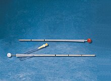
Chest drain
The chest or pleural drainage is used to convey blood, secretions or air from the pleural space (the space between the surface of the lungs and the pleura ) in order to maintain or restore its physiological sub-atmospheric pressure. The chest and pleura ( pleura parietalis ) are opened through an intercostal space and a tube is inserted. A negative pressure can be created using gravity or controlled suction to drain air and / or fluid from the pleural space.
application
A chest tube must be placed in order to restore and maintain the normal sub-atmospheric pressure in the pleural space. This sub-atmospheric pressure is essential for the mechanics of the lungs . Otherwise the lungs could collapse . Atelectasis threatens with less pronounced findings . If the pleural pressure rises, as in tension pneumothorax or severe internal bleeding of the chest cavity, there is a risk of failure of the lungs and heart due to the displacement of the organs and blood vessels in the chest.
The most common use of the drainage is in connection with surgery that requires opening the chest. One or more drains are usually introduced here.
Pneumothorax and / or hemothorax often arise in connection with accidents in which the chest was subjected to greater violence.
In connection with numerous diseases or injuries of the chest cavity and the cardiovascular system can Serothorax , chylothorax , empyema , pneumothorax , hemothorax or hemopneumothorax occur slowly or rapidly.
method
The chest tube is inserted either open, as part of a thoracotomy or thoracoscopy , or "closed" through a small skin incision.
The placement of a chest tube is a surgical procedure in the chest (thorax) , which is usually carried out by surgeons , but must also be mastered by all doctors working in emergency medicine or intensive care as a life-saving immediate measure .
In elective (i.e. plannable, non-time-critical) interventions, the application of a thoracic drainage in the operating room or in the functional department (e.g. endoscopy) is preferable to the location of an intensive care or normal ward for reasons of hygiene.
As a rule, the chest tube is created by means of a 2 to 3 cm incision ( mini-thoracotomy ). After the incision with a scalpel and dissection with scissors, the pleura to be drained is palpated with the finger and loosened. The alternative application of the drainage by puncture with a trocar harbors the risk of injury to the lung tissue and subsequent bleeding.
Chest drains are usually made of silicone or PVC, with X-ray contrast strips that go through the distal eye of the drain, the so-called "Sentinel Eye". In addition to the material, they also differ in size and shape. There are straight and curved chest tubes. Thoracic trocar catheters have only straight shapes. The sizes range from 8 Charrière (CH) to 36 CH . There are basically two different drainage methods: Bülau and Heber . The siphon drainage principle is based on gravity. The thorax level must therefore always be above the drainage container. The Bülau drainage principle (Gotthard Bülau, 1835–1900), on the other hand, generates passive permanent suction, the extent of which can be varied on Bülau's drainage apparatus.
In contrast to the Bülau drainage, the Monaldi drainage (named after Vincenzo Monaldi, 1899–1969, who invented the intrapulmonary cavern suction drainage in 1938) is usually thinner ( smaller-lumen ); it is mainly used for the treatment of a pneumothorax (air between the inner and outer lung membrane ). The smaller lumen is sufficient for air drainage and enables a smaller skin incision with less invasive surgery. The puncture is made on the front of the chest below the collarbone (in the second intercostal space (ICR) in the medioclavicular line). Here, the intercostal space is opened or pierced either by making an incision with a scalpel, sharp scissors or with a trocar.
All forms of thoracic catheters can be used to treat simple sero- or hemothoraxes.
If there is no drainage system available and a dangerous tension pneumothorax is present, it should immediately be converted into an open pneumothorax by puncturing the thorax with several large-lumen cannulas . This relieves the dangerous overpressure and the unaffected lungs are ventilated normally again. Then, under orderly conditions, the chest drain is put on.
Drainage systems
Various technical devices are used to ensure that the negative pleural pressure is maintained and to regulate it.
The simplest and therefore most commonly used in emergency medicine is a Heimlich valve .
Drainage suction for thoracic drainage developed from the one-chamber system (underwater lock and secretion chamber in one chamber) to the three-chamber system (with or without suction). The way in which the most common disposable systems work today is based on the three-bottle system.
Single chamber systems
The first and simplest chest drainage system consisted of a bottle of liquid into which the drainage tube is immersed. The goal of removing both air and secretion from the pleural space and preventing the air from getting back into the pleural space was achieved through this “water lock principle”. During expiration (exhalation), air is mobilized from the pleural space through the water lock. The secretion remains in the water lock. However, no air can get back through the water into the pleura. The one-bottle system was easy to use as long as no large amounts of secretion prevented the outflow and run-in of air and liquids. In single-bottle systems, in which the immersion depth of the riser pipe cannot be changed, the greater the amount of secretion, the greater the resistance in the system, and thus the resistance of the airways. This technology is now obsolete.
Bicameral systems
This form of thoracic drainage consists of the above-mentioned water lock and the secretion collection chamber, in which the secretion is collected without affecting the function of the water lock.
This system must never be operated with active suction, as there is no control over the negative pressure prevailing in the system.
Often the alternating pressure of breathing in combination with a water lock is not enough to allow the pleural space to develop sufficiently again. In this case, a vacuum is required, which then requires the use of a three or, better, four-chamber system.
Three and four chamber systems
The third chamber is used to limit the suction and is used when suction is generated by a pressure transducer or central vacuum that does not have a vacuum meter or with which a low vacuum cannot be set (e.g. for high vacuum regulators with a display up to 1 bar (corresponds to 1000 mbar)).
Filling this additional chamber with water prevents excessive suction from damaging the lungs. If the suction (measured in centimeters of water column) exceeds the weight of the previously filled water column, it is sucked down into an equalization chamber and air can flow in. In this way, the maximum desired suction is always maintained. Typical for such thoracic drainage systems is the constant “bubbling” in the suction chamber, which limits the maximum negative pressure in the system. This is a critical safety factor. The fourth chamber shows changes in pressure in the pleural space.
Electronic drainage systems
In the S3 guideline of the German Society for Thoracic Surgery and the German Society for Pneumology, digital or electronic systems are recommended, as they allow modern drainage management, which shortens the length of stay in hospital and reduces treatment costs. With this type of drainage system, an electronically controlled suction system takes on the task of generating and controlling the vacuum. These systems generally consist of a canister, which replaces the secretion bottle, and an electronically controlled motor unit system. This replaces the vacuum source, suction control bottle and water lock. It is important to know that these systems are not "pumps" that apply a "permanent suction". Rather, the set vacuum value is monitored on the patient, and the system only intervenes if the actual and setpoint values differ. With a drainage time of 2.5 days after an uncomplicated lobectomy , the absolute running time of the device is only around 90 minutes. The first electronic systems were introduced in 2006.
The use of a digital system opens up new possibilities, e.g. B. the course of therapy of the patient can be measured and recorded. At the same time, patient safety is increased, as most digital systems have automated alarms and warnings.
In addition to the miniaturization of the system, which favors the early mobilization of patients, the monitoring electronics with alarm functions as well as the generation of objective data with regard to air leakage and, more recently, fluid measurement for a manufacturer, are important advantages of these systems. With the help of the electronic drainage system, it is possible to monitor the pleural space close to the patient in real time. The measurement is made as close as possible to the pleural space - namely at the connection between the drainage catheter and the hose system. A new feature of this hose system is the fact that on the one hand it is a double lumen hose and on the other hand the geometry in the connecting part to the unit is designed in such a way that air and liquid are separated here. The double lumen hose is used to convey air and liquid. The thinner of the two tubes is used to measure pressure near the pleural space. Studies show, however, that this close measurement to the pleural space provides data that come very close to reality or correspond to it.
Based on data and the possibility of storing, viewing and interpreting them over time - healing is a dynamic process (!) - it was possible to document in studies that the drainage time for anatomical resections can be shortened by one day. Other studies have come to the conclusion that electronic systems do not offer any advantages in terms of length of stay in hospital or length of stay of the chest tube.
The measurement of the air leakage (= alveolo- pleural fistula or bronchopleural fistula) is carried out according to the "blade wheel": Via the speed of the integrated in the system paddle wheel which corresponds to the conveyed quantity of air is, by means of a mathematical algorithm very precise actually delivered air quantity calculated and shown in ml / min on the display. After a running time of one hour, a graphic can also be shown on the display that shows the progress of the leak over time.
Another very important aspect of this measurement is that it provides objective data that does not depend on the observation and interpretation of the people involved in the treatment. It could be shown that by using this system the discrepancies in the assessment of the healing process are significantly smaller compared to conventional systems.
Another advantage of these systems are their monitoring and alarm functions, which increase the safety of treatment and make work easier for the nursing staff in particular. Electronic systems also enable patients to be mobile. This increases the quality of life and has been shown to accelerate recovery.
Single and multi-chamber systems with the water lock principle, electronically controlled suction system, disposable and reusable, belong to the conventional systems. Reusable systems are used less and less, the causes are poor hygiene and problems with tightness after re-sterilization.
Other types of drainage in the chest
Mediastinal drainage
This localization of a drain is mainly used in cardiac surgery . The drainage is behind the breastbone (= sternum ). and placed along the operated heart if necessary. The main focus in this indication is bleeding control. Whether this drainage is provided with active suction depends on various factors such as the preferences and experience of the treating doctor, the individual situation of the patient, etc. Mostly made of very soft silicone with X-ray contrast strips with a diameter of approx. 28 CH. Use after operations on the heart (in combination with pleural drainage) and in the mediastinum . Location: within the mediastinum.
Pericardial drainage
The drainage of the pericardium can be obtained by puncture (transcutaneous) or by open surgery. In the first case, small-lumen drains are used, which are not suitable for a thick effusion ( e.g. hemopericardium ). With pericardial drainage, the drainage is usually guaranteed by gravity.
If a pericardial drainage is surgically inserted (usually subxiphoidal), it is possible to use a larger-lumen drainage, with which the risk of constipation is lower.
history
Already from prehistoric times there is evidence of thoracic interventions such as resection of ribs or pleural functions in pleural empyema . In ancient times, these procedures were further used and improved. In the Corpus Hippocraticum, for example, the puncture of body secretions from the pleural space with hollow tubes made of tin is described. No significant advances in thoracic surgery took place in the Middle Ages. Only Fabrizio D'Aquapendente (1537–1613) described permanent drainage of a pleural empyema with a thread drainage and a special pleural puncture needle with fixation wings. In 1795, the French surgeon Jean Louis Petit advised surgical removal of a hemothorax in the case of penetrating thoracic injuries and, in contrast to the wait-and-see attitude that prevailed at the time, recommended an early puncture. Such recommendations met with bitter resistance in some cases. Guillaume Dupuytren thought punctures or drainages in the thorax area were too dangerous and strictly rejected them because of possible late effects and scarring. After surgery was recognized as a scientific discipline at the beginning of the 19th century, August Gottlieb Richter did pioneering work in the field of thoracic surgery. In addition to operative removal of empyemas and hemothoraces and relief of pneumothoraces, he also performed operations on the mediastinum . He used metallic trocars for postoperative drainage, including drainage of the pericardium. Although the realization gradually gained acceptance that intrathoracic fluid or air accumulations require permanent drainage and not just a single puncture, it was only Gotthard Bülau , internist and senior physician at the Hamburg St. Georg Hospital , who succeeded in reducing the physiological sub-atmospheric pressure in the pleural space after the To restore and maintain surgery. He used a so-called (under) water lock. In 1951, the Italian surgeon Vincenzo Monaldi described a suction technique in which a tube is placed below the middle of the clavicle (medioclavicular) (Monaldi drainage).
Individual evidence
- ↑ Albert Linder: Thoracic drainage and drainage system - modern concepts . UNI-MED Verlag, ISBN 978-3-8374-1442-4 .
- ↑ J. Brokmann et al. a .: Emergency medicine revision course: To prepare for the "emergency medicine" exam. Springer, 2007, ISBN 978-3-540-33702-7 , pp. 105-106. (books.google.de)
- ↑ Thomas Kiefer: Thoraxdrainagen . Springer Verlag, 2016, ISBN 978-3-662-49739-5 , pp. 50, 60 .
- ↑ Christoph Weißer: Thorax surgery. In: Werner E. Gerabek , Bernhard D. Haage, Gundolf Keil , Wolfgang Wegner (eds.): Enzyklopädie Medizingeschichte. de Gruyter, Berlin / New York 2005, ISBN 3-11-015714-4 , p. 1397.
- ↑ DGT, DGP: S3 guideline: Diagnosis and therapy of spontaneous pneumothorax and post-interventional pneumothorax . Ed .: AWMF.
- ↑ G. Miserocchi, D. Negrini: Pleural space: pressure and fluid dynamics . In: RG Crystal, JB West (Ed.): The lung: scientific foundations . tape 2 . Lippincott-Raven Press, New York / Philadelphia, Pa. 1997, ISBN 0-397-51632-0 , Chapter 88, pp. 1217-1225 .
- ↑ José M. Mier, Laureano Molins, Juan J. Fibla: The benefits of digital air leak assessment after pulmonary resection: Prospective and comparative study . In: CIR ESP . tape 87 , no. 6 , 2010, p. 385-389 .
- ↑ Cecilia Pompili, Frank Detterbeck, Kostas Papagiannopoulos, Alan Sihoe, Kostas Vachlas, Mark W. Maxfield, Henry C. Lim, Alessandro Brunelli: Multicenter International Randomized Comparison of Objective and Subjective Outcomes Between Electronic and Traditional Chest Drainage Systems . In: The Annals of Thoracic Surgery . tape 98 , no. 2 , August 1, 2014, p. 490-497 , doi : 10.1016 / j.athoracsur.2014.03.043 , PMID 24906602 .
- ↑ Gonzalo Varela, Marcelo F. Jiménez, Nuria Maria Novoa, José Luis Aranda: Postoperative chest tube management: measuring air leak using an electronic device decreases variability in the clinical practice . In: European Journal of Cardio-Thoracic Surgery . tape 35 , no. 1 , January 1, 2009, p. 28-31 , doi : 10.1016 / j.ejcts.2008.09.005 .
- ↑ RJ Cerfolio, AS Bryant: The quantification of postoperative air leaks . In: Multimedia Manual of Cardiothoracic Surgery . 2009, doi : 10.1510 / mmcts.2007.003129 .
- ↑ AL McGuire, W. Petrich, DE Maziak, FM Shamji, SR Sundaresan, AJE Seely, S. Gilbert: Digital versus analogue pleural drainage phase1: prospective evaluation of interobserver reliability in the assessment of pulmonary air leaks . In: Interactive CardioVascular and Thoracic Surgery . tape 21 , no. 4 , 2015, p. 403-408 , doi : 10.1093 / icvts / ivv128 , PMID 26174120 .
- ↑ D. Dani sealed: Benefits of digital thoracic drainage system. In: Nursing Times . tape 108 , no. 11 , March 13, 2012, p. 16-17 ( nursingtimes.net ).
- ↑ Stefan J. Schaller and a .: Early, goal-directed mobilization in the surgical intensive care unit: a randomized controlled trial . In: The Lancet . tape 388 , no. 10052 , 2016, p. 1377-1388 .
- ↑ a b c d Annegret Gahr, Ralf Gahr: The history of thorax traumatology. In: Ralf Gahr (Ed.): Handbook of Thorax Traumatology. Volumes I and II, Einhorn-Presse Verlag, 2007, ISBN 978-3-88756-812-2 .
- ^ V. Monaldi, F. De Marco: Terminal technic of the suction drainage of pulmonary caverns . In: Munch Med Wochenschr . 1950.
- ^ K. Knobloch: A tribute to Gotthard Bulau and Vincenzo Monaldi . In: Interact Cardiovasc Thorac Surg . tape 7 , no. 6 , December 2008, pp. 1159 , doi : 10.1510 / icvts.2008.181750A .


