Inguinal hernia
| Classification according to ICD-10 | |
|---|---|
| K40 | Inguinal hernia |
| ICD-10 online (WHO version 2019) | |


A hernia , even inguinal hernia (Latin inguinal hernia called), is a hernia in the area of the inguinal canal . Muscles, tendons and connective tissue form the firm outer covering of the body cavities, such as the abdomen. There are natural "weak points" on this shell. One of the most important is where the spermatic cord enters the abdominal wall. If there is an enlargement here, tissue from the abdominal cavity escapes into the inguinal canal. A "break" (hernia) occurs. The inguinal hernia is the most common hernia alongside the umbilical , femoral and incisional hernias . It occurs in men and women of all age groups in a ratio of men: women = 9: 1. Every year around 0.5% of the population develop an inguinal hernia. In childhood, it occurs in one to three percent of all children, in premature babies in around five percent. In most cases, the inguinal hernia is treated surgically. A hernia is operated on if it causes pain or if there is a risk of entrapment with the life-threatening death of parts of the intestine. Inguinal hernias can also be associated with erectile dysfunction ("impotence"). In other mammals as well , inguinal hernias occur predominantly in male individuals, especially after open castration .
anatomy
Inguinal hernias occur in the inguinal canal . Along with three nerves (genital ramus of the genitofemoral nerve , the ilioinguinal nerve and the iliohypogastric nerve ) and lymph vessels, the spermatic cord ( funiculus spermaticus ) in male mammals and the cervical ligament ( ligamentum teres uteri ) run in this. Reaches the hernial sac to the male in the scrotum ( scrotum ), one speaks of a scrotal hernia or hernia ( Hernia scrotalis or Skrotalhernie ), which is a special form of the hernia.
The inguinal canal has an entrance and an exit:
- the outer inguinal ring (annulus inguinalis superficialis), as the exit of the inguinal canal;
- the inner inguinal ring (annulus inguinalis profundus), as the entrance into the inguinal canal from the abdominal cavity.
The outer inguinal ring is a slit-shaped opening in the fascia (muscle skin) of the external oblique abdominal muscle (Musculus obliquus externus abdominis). The inner inguinal ring lies between the free edge of the inner oblique abdominal muscle (Musculus obliquus internus abdominis), the straight abdominal muscle (Musculus rectus abdominis) and the inguinal ligament (Arcus inguinalis, also ligamentum inguinale) .
The inguinal canal is limited:
- front: aponeurosis of the externus abdominis oblique muscle;
- back: Fascia transversalis abdominis;
- Roof: Musculus transversus abdominis and parts of the musculus obliquus internus abdominis;
- Floor: inguinal ligament.
classification
According to the location of the hernial port (opening for the hernial sac), a distinction is made between direct ( medial ) and indirect ( lateral ) inguinal hernias. The term "direct" refers to the fact that the break runs directly through the posterior wall of the inguinal canal (and not through the inner inguinal ring), and medially to the position in the center of the epigastric vessels. Correspondingly, indirect inguinal hernias get through the inner inguinal ring into the inguinal canal and lie to the side (“lateral”) of the epigastric vessels. The division of inguinal hernias into these two forms makes a significant contribution to understanding the disease, but is of little importance for modern inguinal hernia surgery. A modern classification was developed by Nyhus and has proven itself most effective for study questions to this day.
Symptoms and their causes

In childhood, the inguinal hernia arises through a connecting channel, the processus vaginalis testis (also Proc. Vaginalis peritonei , "vaginal skin process"). This is a part of the peritoneum that is everted into the scrotum through the inguinal canal in the course of the fetal development by migration of the testicle developing in the abdominal cavity into the scrotum. This channel usually closes at birth. If this does not occur, abdominal organs can penetrate into the canal. Congenital hernias are therefore always indirect (lateral) hernias.
In adulthood, in addition to a predisposition in the sense of an abdominal wall weakness or an excessively wide inguinal canal, an increase in internal abdominal pressure, for example due to hard physical work, chronic coughing , strong pressure in the case of chronic constipation, can trigger the formation of a hernia. In addition, fractures often occur in women during pregnancy . Hard work alone is not recognized (and compensated) as the cause of an inguinal hernia.
The symptoms of an inguinal hernia can include:
- in childhood: mostly painless swelling in the groin, which is often discovered by chance while changing diapers or while taking care of the body.
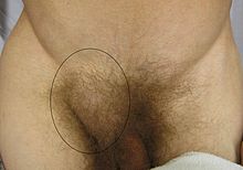
- In adulthood: mostly visible or palpable swelling in the groin area, which can be provoked by physical exertion, straining or coughing. When lying down, you can usually “push away” the “bump” without any problems. Severe pain is by no means the rule, but mostly just a certain feeling of pressure. With gradually increasing enlargement, it can also become evident in men as a swelling or enlargement of the scrotum (testicular fracture).
- Frequent testicular pain is caused by irritation of the genital branch in the inguinal canal .
- Pain in the upper part of the adductors on the inside of the thigh is also possible.
- Sudden severe pain in the groin area with swelling that cannot be pushed away in the groin area is a pinched break. Quick action is required here and a surgeon must be consulted immediately.
Fractures are particularly dangerous if organs of the abdominal cavity - for example parts of the intestine - remain trapped in the fracture ( incarceration ). If this rare complication occurs, severe pain occurs in the area of the hernia and the entire abdomen, the character of which differs from the symptoms associated with an uncomplicated inguinal hernia. If the entrapment of a fracture is recognized in good time, it may be possible to reduce it . As a result of the entrapment, the entrapped intestine swells and thereby constricts itself from the blood supply. The break can then no longer be pushed away. This is known as incarceration and strangulation can lead to the death ( necrosis ) of the trapped organ. It can also lead to an ileus (bowel obstruction). Both situations are life-threatening and require immediate surgery. A part of the intestine may then have to be removed ( resection ).
Fractions tend to get bigger and bigger over time.
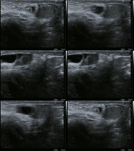
Sonography is helpful for the exact diagnosis of small hernias. The MRI is only suitable as a dynamic MRT to detect a break. However, a negative test result does not definitely rule out a hernia. Most important symptoms of an inguinal hernia: Pain when lifting, coughing or pressing the abdominal press, which decreases when lying down, should suggest a hernia. In addition to an inguinal hernia, other diseases also cause groin pain. If there is no inguinal swelling and a negative ultrasound, one should think of hip diseases - in athletically active patients - diseases of the pubic bone , adductors, hip flexor and lower spine . Urological diseases must be excluded in these cases, as well as abdominal diseases ( divertculosis , appendicitis ) or urological diseases.
history
Inguinal hernias are believed to have been around since 2000 BC. Known. For a long time, however, there was a lack of knowledge of the basics, the origin and successful treatment of inguinal hernias. The treatment methods have been dangerous for centuries and often with disastrous results. A surgical therapy with removal of the hernial sac while protecting the testicle was first described by Celsus in the 1st century AD. Later the surgeon, Ritter and Count Hans von Toggenburg (around 1477) and the surgeon Caspar Stromayr (around 1559) were known for their surgical treatment of hernias , both of whom were also ophthalmologists. The operation of a pinched hernia was first described in 1556 by the French surgeon Pierre Francou . In the statutory accident insurance of 1884, it was initially disputed whether hernias could be compensated as work-related accidents. It was not until 1890 that the results of the surgical treatment were decisively improved by the pioneering work of Edoardo Bassini of the Royal University of Padua. Since then, numerous modifications and new methods have been introduced into therapy.
Conservative care
To this day, a conservative treatment option is considered to be a hernia ligament . With the correct technical adjustment, complications can be avoided. This form of care is used today for patients who can no longer be operated on due to their age or other medical circumstances.
Furthermore, especially younger people who do heavy physical work can be treated with a hernia ligament after the diagnosis of an inguinal hernia up to the date of surgery. This is useful if a prompt surgery is not possible.
The operative closure
The only way to avoid entrapment of organs and thus serious or life-threatening consequences is surgical closure. With around 230,000 surgical interventions, the inguinal hernia is one of the most frequently surgically treated diseases in Germany.
The surgeon Dieffenbach is said to have said that "one shouldn't let the sun go down over a trapped fracture".
A distinction is made between open surgical procedures and minimally invasive procedures (“keyhole method”).
Open procedure (herniotomy)
The open method goes back above all to Edoardo Bassini (1890), whose principle consists in closing the hernial port and reinforcing the inguinal canal wall using a specific suture technique. About a hundred years later, this technique was superseded by the inguinal hernia operation developed by the Canadian doctor Edward Earle Shouldice and named after him , which follows the same principle, but uses a modified suture technique and is particularly indicated for smaller hernial orifices and younger patients.
The open implantation of plastic meshes, which can consist of absorbable and non-absorbable or titanium-coated components and are usually introduced using the Lichtenstein technique of the American surgeon Irving L. Lichtenstein, is also widespread .
In childhood, the hernial sac is sought out in the groin, the contents - if present - pushed back into the abdomen, the hernial sac is then removed and closed. A reinforcement of the posterior wall of the inguinal canal as indicated above is not carried out in the case of a child's inguinal hernia. Foreign material is also not brought in, as it does not grow with you.
Minimally invasive procedures
With minimally invasive techniques, the hernial port is always closed with a mesh. Here, again, a distinction is made between two methods:
On the one hand , using the so-called TAPP technique, the mesh can be placed laparoscopically - that is, via a laparoscopy from the abdomen - over the hernial orifice. It is necessary to go into the abdominal cavity, the peritoneum has to be cut open and closed again at the end of the operation. The mesh is fixed using metal clips, absorbable clips, sewing on or using tissue adhesive. With the TEP technique, the mesh is also placed over the hernial port via minimally invasive access after carefully pushing the layers of the abdominal wall apart. The mesh is placed between the peritoneum and the muscles without any further fixation. In the minimally invasive procedure, no foreign material (mesh) is introduced into the groin during childhood , as this cannot grow with the patient. The hernial port visible by laparoscopy is closed with a suture.
Pros and cons of the procedures
Each of the procedures mentioned has its strengths and weaknesses. In principle, one cannot say that one of the techniques is in principle the superior or safer method. In addition, the groin encompasses an extremely sensitive area of the body, in which the central nerve tracts (or in men also the spermatic cord) run and thus surgical interventions are associated with an additional risk. Various studies point to the relatively high number (20%) of postoperative complaints, from “nerve irritation, paresthesia, pain and restricted mobility” to persistent discomfort during sexual intercourse and even infertility. However, there are also indications that such an operation with the installation of a mesh can improve pre-existing disorders of sexual functions. Affected patients should seek intensive advice on which surgical procedure and which material (plastic, titanized or oxygen-coated mesh) to use.
The procedures that cover the hernial gap with a mesh (both open and closed) are referred to as "tension-free" procedures and should be immediately resilient and have a lower recurrence rate than the Shouldice method with larger hernial openings. Mesh implants, depending on the material (plastic or titanized surfaces), lead to different desired or undesired scarring, which in turn can result in neuralgia (nerve pain). Minimally invasive techniques are mostly perceived by patients as less painful in the early recovery phase and are therefore particularly indicated for bilateral operations in one session. In the late phase, however, painful conditions that are difficult to treat are occasionally observed, which may be due to metal clips with which the mesh is fixed to prevent slipping, especially with the TAPP technique. No metal clips are used with TEP technology. Metal clips are still predominantly used in TAPP technology. Some surgeons have recently started using absorbable clips or fibrin glue instead to prevent nerve damage. With modern mesh implants, which are coated with a very thin layer of titanium, there is a hydrophilic connection with the tissue, so that the problematic fixation can usually be dispensed with. In contrast to the TAPP technique, the TEP technique does not require an operation in the abdominal cavity with cutting open and sewn again, as the minimally invasive operation is only performed between the layers of the abdominal wall. There is therefore no risk of injury to the internal abdominal organs with the TEP technique. Cases are occasionally described here for the TAPP technique. Especially with previous operations in the abdominal cavity with the corresponding adhesions , the risk of injury is significantly increased with the TAPP technique. Another good indication for the use of minimally invasive techniques is the operation of recurrent hernias, that is, of inguinal hernias that were previously operated on openly but have now reappeared.
Basically, if a recurrent hernia occurs after a minimally invasive operation , an open surgical technique should be chosen. If the patient has already been operated on with an open surgical procedure, a minimally invasive technique should be used.
The change in surgical technique is intended to reduce the complication rate of a new operation, which is increased due to existing scarring.
Open procedures can mostly be performed under local anesthesia. In this way, risks of anesthesia can be avoided. The open techniques are also particularly suitable for outpatient operations. But also minimally invasive operations are increasingly being carried out on an outpatient basis using routine technology. The choice of procedure should always be made individually.
In childhood, the discussion between open and minimally invasive procedures has not yet been decided.
Differential diagnosis
The most common symptom, severe swelling, can also be attributed to a femoral hernia , hydrocele, or varicocele . Furthermore, enlarged lymph nodes can cause swelling.
literature
- Edmund Andrews: A history of the development of the technique of herniotomy. In: Annals of medical history. New series, 7, 1935, pp. 451-466.
- Paul Koch: The history of the herniotomy except for Scarpa and A. Cooper . Dissertation, Berlin 1883.
- Michael Sachs, Albrecht Encke : The repair procedures of inguinal hernia surgery in their historical development. In: Zentralblatt für Chirurgie. Volume 118, 1993, pp. 780-787.
- Caspar Stromayr: Practica copiosa from the right ground of the Bruchschnidts (1559). Edited by Werner Friedrich Kümmel together with Gundolf Keil and Peter Proff, Munich 1983.
- LM Nyhus: Classification of groin hernia: milestones. In: Hernia. Volume 8, Number 2, May 2004, ISSN 1265-4906 , pp. 87-88, doi: 10.1007 / s10029-003-0173-6 , PMID 14586776 (review).
- S1 guideline inguinal hernia, hydrocele of the German Society for Pediatric Surgery (DGKCH). In: AWMF online (as of 2010)
Web links
- LM Nyhus: Classification of groin hernia: milestones. In: Hernia: The journal of hernias and abdominal wall surgery. Volume 8, Number 2, May 2004, pp. 87-88, doi: 10.1007 / s10029-003-0173-6 , PMID 14586776 (Review) (On the classification of inguinal hernias).
- www.wissenschaft.de: Information on minimally invasive inguinal hernia surgery as an alternative to open surgery
- E-learning on inguinal hernias with images and videos
Individual evidence
- ↑ Jens Krüger: The inguinal hernia - operative or conservative treatment? Ed .: Jens Krüger. Sports Surgery Edition, Columbia SC February 10, 2018, p. 14th ff .
- ↑ Jürgen Zieren, Charalambos Menenakos, Marco Paul, Jochen M. Müller: Sexual function before and after mesh repair of inguinal hernia. In: International Journal of Urology. Volume 12, No. 1, January 2005, pp. 35-38, doi: 10.1111 / j.1442-2042.2004.00983.x
- ↑ Duale Series Anatomie, 2nd edition, p. 281.
- ↑ a b Jens Krüger: The inguinal hernia - operative or conservative treatment? Ed .: Jens Krüger. Sports Surgery Edition, Columbia SC February 10, 2018, p. 26 .
- ↑ Jens Krüger: The inguinal hernia - operative or conservative treatment? Ed .: Jens Krüger. Sports Surgery Edition, Columbia SC February 10, 2018, p. 33 .
- ↑ Jens Krüger: The inguinal hernia - operative or conservative treatment? Ed .: Jens Krüger. Sports Surgery Edition, Columbia SC February 10, 2018, p. 27-30 .
- ^ A. Weir, P. Bruckner, E. Delahunt et al .: Doha agreement meeting on terminology and definitions in groin pain in athletes . In: British Journal of Sports Medicine . tape 49 , 2015, p. 768-774 , PMID 26031643 (English, bmj.com [accessed May 21, 2018]).
- ↑ Christoph Weißer: Hernias. In: Werner E. Gerabek , Bernhard D. Haage, Gundolf Keil , Wolfgang Wegner (eds.): Enzyklopädie Medizingeschichte. De Gruyter, Berlin / New York 2005, ISBN 3-11-015714-4 , p. 574.
- ↑ Gundolf Keil: "blutken - bloedekijn". Notes on the etiology of the hyposphagma genesis in the 'Pommersfeld Silesian Eye Booklet' (1st third of the 15th century). With an overview of the ophthalmological texts of the German Middle Ages. In: Specialized prose research - Crossing borders. Volume 8/9, 2012/2013, pp. 7–175, here: pp. 10 f.
- ↑ Barbara I. Tshisuaka: Francou, Pierre. In: Werner E. Gerabek u. a. (Ed.): Encyclopedia of medical history. 2005, p. 419 f.
- ↑ Collection of sources on the history of German social policy from 1867 to 1914 , Section II: From the Imperial Social Message to the February Decrees of Wilhelm II (1881–1890) , Volume 2, Part 2: The expansion legislation and the practice of accident insurance , edited by Wolfgang Ayass . Darmstadt 2001, pp. 1051, 1068, 1070, 1081-1085, 1215-1217, 1227-1229, 1231, 1287.
- ↑ Schöne, Scheuerlein and Settmacher: Diagnosis and treatment of inguinal hernias. (PDF) In: MMW - Advances in Medicine Volume 151, 2009, pp. 44–49.
- ^ Ferdinand Sauerbruch, Hans Rudolf Berndorff: That was my life. Kindler & Schiermeyer, Bad Wörishofen 1951; cited: Licensed edition for Bertelsmann Lesering, Gütersloh 1956, p. 315.
- ↑ Jens Krüger: Glossary - Explanation of the medical term. In: website. Jens Krüger, May 21, 2018, accessed on May 21, 2018 .
- ↑ Jens Krüger: The inguinal hernia - operative or conservative treatment? Publisher = Edition Sports Surgery . Ed .: Jens Krüger. Columbia SC February 10, 2018, p. 47-54 .
- ↑ F. Schenten: Risk factors for the development of inguinal hernia recurrence - A retrospective 10-year analysis. (PDF; 1.6 MB) Dissertation, RWTH Aachen, 2008.
- ↑ Veronika Hackenbroch: knife into the room . In: Der Spiegel . No. 5 , 2009, p. 104-106 ( online ).
- ↑ J. Grace, C. Menenakos, M. Paul, JM Müller: sexual function before and after mesh repair of inguinal hernia. In: International Journal of Urology. Volume 12, Number 1, January 2005, pp. 35-38, doi: 10.1111 / j.1442-2042.2004.00983.x , PMID 15661052 .
- ^ DocCheck Medical Services GmbH: Inguinal hernia. Retrieved August 25, 2019 .
