Cerebral cortex
The cerebral cortex (Latin cortex cerebri , short: cortex ) is the outer layer of the cerebrum ( telencephalon ) rich in nerve cells (neurons ).
Although the Latin word cortex translated simply means bark 'and Cortex (or cortex ) actually the entire cortex called, is cortex in the jargon also used restrictive of the cerebral cortex. Accordingly, the adjective means cortical (or cortical ) actually "the entire cortex on" but is often used "the cerebral cortex on" in the strict sense of.
Depending on the region, the cerebral cortex is only 2 to 5 millimeters thick and part of the gray matter ( substantia grisea ) of the cerebrum. The nerve fibers of the neurons of the cerebral cortex run below the cerebral cortex and are part of the white matter ( substantia alba ) of the cerebrum, which is also referred to here as the medullary bed . The cerebral cortex and medullary beds together form the cerebral mantle ( pallium cerebri ). Below the cerebral cortex, subcortically , there are further sections of gray matter of the cerebrum as subcortical core areas ( basal ganglia , claustrum and corpus amygdaloideum ).
structure
Macroscopic breakdown
The terminology of the cerebral lobes and convolutions still valid today was proposed in 1869 by Alexander Ecker (1816–1887).
Lapping
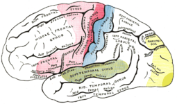
The cortex can be roughly divided into five to six lobes ( lobi ), which are separated from one another by deeper fissures ( fissures ). Of these are on the brain surface:
- Frontal lobes or frontal lobes ( lobus frontalis )
- Parietal lobe or parietal lobe ( lobus parietalis )
- Occipital lobe or occipital lobe ( lobus occipitalis )
- Temporal lobe or temple lobe ( lobe temporalis )
Covered by parts of the frontal, parietal and temporal lobes lies the
- Island lobes ( lobus insularis )
In addition, some authors summarize certain developmentally older parts of the cortex (e.g. cingulate gyrus and hippocampus ) as sixth
- Limbic lobes ( Lobus limbicus )
The subdivision of these flaps is not only morphologically but also functionally important, since each flap has a special primary processing area:
- There are various areas in the large frontal lobe, the most important of which are the motor centers of the cerebrum in and around the precentral gyrus . In the rostral (anterior) sections lies the prefrontal cortex , which is associated with action planning and initiation. In addition, basic characteristics of personality seem to be localized here.
- The parietal lobe, in which the primary sensitive center ( gyrus postcentralis ) is located, joins the posterior .
- The visual center ( area striata ) is located at the pole of the occipital lobe
- On the inside of the temporal lobe is the hearing center ( area temporalis granulosa ) in the so-called Heschl's transverse turns ( gyri temporales transversi )
- The island bark is the least explored. Among other things, this is where the primary taste cortex is located . It is believed that this is also the primary center for basal viscerosensitivity (information from the intestines).
- In the limbic lobe (also limbic system ), old interconnection patterns are processed, the most prominent representatives of which are memory functions and emotional processes.
Convolution ( gyration )
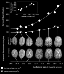
In many mammals, the cerebral cortex is characterized by numerous turns (Latin-Greek gyri , singular gyrus ), crevices (Latin fissurae , singular fissura ) and furrows (Latin sulci , singular sulcus ). The folding serves to enlarge the surface: in humans this is about 1800 cm². When it comes to furrowing the cortex, a distinction is made between a primary furrow, which is approximately the same in all individuals, from a secondary and tertiary furrow, which can be as individual as a fingerprint .
Corrugated brains are called gyrencephalic . In some small mammals (e.g. rodents , hedgehogs ) and birds , the cortex has no furrows ( lissencephalic brain ). The Trnp1 gene has the ability to influence gyration through different expression levels and even to induce it in normally lissencephalic brains.
The lobi and gyri are separated from one another by the fissures and sulci. Their most important representatives are:
- Fissura longitudinalis , which forms the gap between the two hemispheres. The falx cerebri protrudes into the fissura longitudinalis.
- Sulcus centralis separates the frontal and parietal lobes ( gyrus praecentralis or gyrus postcentralis ) and thus the primarily motor from the primarily sensitive cortical field
- The lateral sulcus (also called the Sylvian fissure) lies above the insula and separates the temporal lobe from the frontal and parietal lobes above it
- Sulcus parietooccipitalis between the parietal and occipital lobes
- Sulcus calcarinus divides the primary visual cortex within the occipital lobe into an upper and lower part, which represents the opposite visual field, i.e. above the sulcus calcarinus the lower visual field, below the sulcus calcarinus the upper one.
Histological classification
The cortex can be divided into two categories. On the one hand, due to its histological fine structure in a six-layer isocortex and a three- to five-layer allocortex . Within the cortex shapes, variations in the histological fine structure can be determined, according to which the human cerebral cortex was divided into 52 areas by Korbinian Brodmann in 1909 ( Brodmann areas or fields). Another consideration is the phylogenetic age of the cerebral cortex, after the cortex is divided into a newer neocortex and the older archicortex and paleocortex .
The histological structure of the isocortex is described below. Information on the archicortex can be found e.g. B. under the hippocampus .
Cell types of the cerebral cortex
The six layers of the cerebral cortex are characterized by the presence of certain types of cells. Many of these cells are interneurons , the extensions of which do not leave the cortex and mainly connect GABAerg ( calbindin- positive cells) between cortical neurons . The cortex is characterized by two types of neurons that are histologically related to one another ( calmodulin kinase II-positive cells), presumably arising from the same progenitor cells. One type includes the so-called pyramidal cells , and the other type the granule cells , sometimes referred to as modified pyramidal cells in German-speaking countries , in Anglo-American usage also called thorny star cells .
- Pyramidal cells are the largest cells in the cortex and are named after the triangular cross-section of their cell body, the base of which is usually parallel to the surface of the cortex. They usually have a basal axon directed towards the medullary as well as several basal and one apical dendrite with thorns . The pyramidal cell is the efferent cell of the cerebrum. She is CaMK II positive and uses glutamate as a neurotransmitter . Your axon can be of different lengths; in the case of the particularly large Betz giant cells of the motor cortex, it extends into the spinal cord.
- Granule cells ( thorny stellate cells or modified pyramidal cells ) have a more rounded cell body and many thorny dendrites, one of which emerges apically from the cell and the others begin almost anywhere on the cell body, which is why it is reminiscent of a star in the histological picture. They are also glutamatergic and CaMK II-positive and represent the afferent cells of the cortex, which receive information from other brain areas and especially from the thalamus .
- Interneurons are numerous in the cerebral cortex. They have different forms and are mostly GABAergic and calbindin-positive. Their processes serve to connect nerve cells within the cortex and do not leave it. They are in double Busch cells , Candelaberzellen , unbedornte stellate cells , Fusiform cells , Marinotti cells , horizontal cells and bipolar cells differentiated.
In addition to the nerve cells, the cortex also contains a large number of glial cells . They form the binding substance between the neurons and perform various special tasks for which they are each specialized:
- Oligodendrocytes form the myelin sheaths around the axons
- Protoplasmic and fibrillar astrocytes support the neuronal tissue, form the blood-brain barrier ( Membrana gliae limitans perivascularis ), as well as the blood-liquor barrier on the brain surface ( Membrana gliae limitans superficialis ) and take on numerous functions for the nutrition and maintenance of the neurons
- Microglia are the defense cells of the central nervous system
There is no significant intercellular matrix in the brain, this also applies to the cerebral cortex. The gap between nerve and glial cells is only 10 to 50 nanometers wide.
lamination
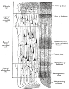
Due to the presence of the different cell types, the cortex can be divided into different layers
From the outside in, these are in the isocortex:
- Lamina I ( stratum molecular ): It is a cell-poor part of the cortex, in which mainly fibers and isolated interneurons are to be found. In embryonic development, this layer is the first to arise, as the first neurons ( Cajal-Retzius cells ) are deposited here, which, however , undergo apoptosis later in development . The other layers develop inversely: layer VI lies under layer I, through which the neurons of layer V then migrate, then layer IV and so on, until layer II is applied to the end. In lamina I there is a pronounced bundle of fibers, the Exner stripe .
- Lamina II ( Stratum granulosum externum ): Here are mainly smaller spiny stellate cells.
- Lamina III ( Stratum pyramidale externum ): This is where smaller pyramidal cells and a strand of fibers, the Kaes-Bechterew strip, lie .
- Lamina IV ( Stratum granulosum internum ): In this layer lie the larger primary afferent spiny stellate cells, which contain projections from other brain areas. Accordingly, this layer is particularly pronounced in the auditory, visual and sensory cortex ( granular cortex ), while it is practically completely absent in motor cortical areas ( agranular cortex ). In the lamina IV there is another fiber network, the outer ligament of Baillarger . In the visual cortex, this band is so strongly developed that it is macroscopically visible to the naked eye. This one as Gennari - or Vicq d'Azyr strips designated strip owes the visual cortex her name as area striata .
- Lamina V ( Stratum pyramidale internum ): It is the seat of the large pyramidal cells that project out of the cerebral cortex. In the motor cortex there are particularly large pyramidal cells ( Betz's giant cells ), while the layer in the granular cortex is completely absent. Furthermore, the deepest of all fiber braids of the cortex, the inner ligament of Baillarger, lies here .
- Lamina VI ( Stratum multiforme ): There are many different pyramidal and thorny stellate cells, as well as numerous interneurons.
In addition to the horizontal layers, the cortex is often organized vertically in columns. These cortical columns are particularly pronounced in the primary sensory areas and are characterized by a strong connectivity within a column. They presumably represent the elementary processing units (modules) of the cerebral cortex.
In a large-scale study, Chen et al. (2008) the modularity of a cortical network consisting of a total of 54 brain regions. In the course of the statistical analysis of the collected data, it could be shown that there are a large number of connections between the regions examined and that the thickness of the cortex alone can provide the necessary information about the modularity of the brain.
Internal organization
There are three different types of axons :
- Association fibers connect different areas within a hemisphere. A distinction is made between short association fibers that connect one gyrus to the next ( fibrae arcuatae ), medium-length (including the Fasciculus arcuatus and Fasciculus uncinatus ) and long association fibers (especially the capsula externa , Fasciculus longitudinalis superior and Fasciculus longitudinalis inferior )
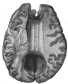
- Commissure fibers connect corresponding areas of opposite hemispheres. Important commissures are the corpus callosum , the commissura anterior , commissura posterior and the fornix ( commissura fornicis ).
- Projection fibers connect the cortex with lower lying areas. The most prominent projection trajectories are the internal capsule as part of the pyramidal tract and the medial lemniscus .
In macroscopy, these different pathways are clearly organized in the medullary bed of the cerebrum. From the outside to the inside, you can see short association fibers ( capsula extrema ), long association paths ( capsula externa ) and, on the very inside, the projection fibers of the capsula interna . The fibers in the cortex have the same arrangement in terms of length and type. If you realize that layers II and IV are afferent and layers III and V are efferent, it is quite logically understandable how the cortex is organized internally:
- Short and medium-length association fibers start in layer III and end in layer II
- Short commissure fibers start in layer III and end in layer II
- Long association and commissure fibers start in layer V and end in layer IV
- Long afferent projections end in Layer IV
- Long efferent projections start in layer V.
- Connections from allocortical areas (olfactory brain, claustrum , amygdala etc.) as well as projections from unspecific thalamic nuclei end in layer VI and partly in layer I.
Functional structure
Local breakdown
In the cerebral cortex there are functional centers that are closely related to the Brodmann areas . The most important functional centers are the primary motor and primary sensory areas.
- The primary motor area, which is part of the motor cortex , is located in the precentral gyrus (Brodmann area 4).
- The primary somatosensitive cortex is located next to it in the postcentral gyrus (areas 1 to 3).
- The primary visual cortex in the occipital lobe forms the most caudal (rearmost) pole of the brain (area 17).
- The primary acoustic cortex is found in the transverse temporal gyri (area 41).
In addition to the primary areas, there is usually a whole series of secondary areas that also exclusively process information from one modality (vision, hearing, motor skills).
These cortex regions play a central role in the processing and awareness of neural impulses, but must not be viewed in isolation, as the entire nervous system represents a multi-interconnected network. The rest of the cerebral cortex is occupied by the association cortex, i.e. areas that receive multimodal input and often have neither clearly sensory nor clearly motor tasks. Today we know that complex skills such as motivation , attention , creativity , spontaneity and, for example, the internalization of social norms depend on them.
Connectome
The connectome is functionally structured according to connected neurons, local structures are irrelevant. In the local subdivisions, the functional parts consist of nerve tissue, i.e. of mixed neurons and glial cells. The connectome so far only describes the connected neurons. In this arrangement, the astrocytes are located between the connected neurons.
Interconnection
The cerebral cortex receives its afferent information mainly from the thalamus . This information includes sensory perceptions of the various sense organs . Areas that receive such information are called sensory areas or projection centers, e.g. B. the visual cortex. The two hemispheres (left and right) receive the information from the other half of the body , as the supply lines cross over to the opposite side. The parts of the cerebral cortex that obtain information from the thalamus are called the primary sensory areas.
Other areas receive impulses from the primary sensory areas and combine information from different sensory organs. These associative areas take up a lot of space in all primates , especially humans.
Finally, the association areas forward information to the motor areas. This is where the commands for all arbitrarily controllable body functions arise and are passed on to the periphery via the pyramidal orbit as the main output of the cerebrum. Parts of the motor cortex are closely interconnected with the basal ganglia and the cerebellum .
In addition to the information that reaches the cortex from the sensory organs via the thalamus, all areas of the cortex receive additional "unspecific" excitations from the thalamic core areas of the reticular formation . These excitations of the ascending reticular activation system (ARAS) are rhythmical, their frequency changing with the degree of vigilance. The spectrum ranges from around 3 Hz in deep sleep and anesthesia to around 40 Hz in wide-awake tension, e.g. B. when reading.
The oscillations of the ARAS are generated in a loop-shaped line between the thalamus and the basal ganglia (striatum, pallidum, caudate nucleus, putamen); they form the natural “ brain pacemaker ”. Electronic brain pacemakers, which were developed in recent years for the treatment of Parkinson's disease , try to replace this activating and inhibiting function of the ARAS.
The evolution and function of the cerebrum
The human brain is not a new development of nature . Like all other organs, it evolved from simple forms. The nervous system develops from a very simple structure, the outer germ layer ( ectoderm ). It is easy to understand that an organ of information processing arises from the outer boundary layer, because this is where the stimuli from the environment hit. It was only in the course of evolution that the sensitive nerve associations were relocated deeper into the neural tube because they are better protected there. The connections to the outside world remained through the now specialized sense organs.
With the development of specialized sensory organs, the formation of a nerve center is connected, which can control the whole body uniformly according to the sensory impressions. Because eyes, ears and chemical senses (taste, smell) develop early in the history of vertebrates, the brain of all vertebrates is designed in the same way for the central integration of these senses.
The endbrain was initially the processing center for the olfactory organ. Because the sense of smell is a general warning and stimulus system of high sensitivity, but says little about the spatial situation or the location of the stimulus source, a connection with the optical and acoustic centers of the midbrain is necessary for the olfactory brain , with which all sensory qualities are shared Level to be united.
This common level already arises in reptiles from an expansion of the endbrain as a telencephalon or rudimentary cortex. Even in frogs and salamanders, this brain structure is designed to integrate the various types of stimuli. For switching the visual, tactile and auditory world from the midbrain to the end brain, a part of the forebrain that develops diencephalon . The thalamus arises from this and sends the specific signals of the midbrain from several core groups to specific regions of the cerebral cortex. This arrangement is called a projection system, the anatomists called the thalamus the "gateway to consciousness".
With the elimination of the scales of fish and the horny scales of reptiles, the whole skin of mammals became a sensitive sensory organ, which also comes into holistic connection with the other sensory qualities via projection tracks in the cortex.
A nerve center, in which all the qualities of the environmental signals are brought together, would not make sense if no commands for the reactions of the organism could be formed in it and passed on to the executive organs. Because the olfactory organ has a controlling access to complex behaviors right from the start, the olfactory brain, which has been expanded to become the center of integration of all senses, can fall back on these control paths in order to develop holistic behavioral steps from the unification of all sensations.
This integration of the neocortex, which connects all the senses to a whole and creates meaningful behavior patterns from it, already enables rats , cats etc. to behave intelligently that does not occur in insects or simple organisms. This shows that even birds and mice can use their integrative center, the cerebral cortex, not only as a command center, but also as a particularly powerful information store (memory) (see also: Birds' brain and cognition ). A fly never learns to avoid colliding with a window pane, while after some experience a bird learns to be careful with the transparent wall.
Only animals that have a cortex can be trained, that is, they develop a memory for verbal instructions, which can also dominate the innate behavioral patterns. This learning ability is clear in the dolphins , which, as mammals, have a powerful cortex and are easy to train, while the relatively cerebral sharks are not very suitable for training.
With the development of the cortex, a playful phase of the young animals increasingly emerges, which is to be understood as the learning phase of the cerebral cortex and gives us the impression that these animals (e.g. dogs, cats, etc.) feel similar mental states as humans .
A powerful development of the cerebral cortex was triggered in the monkeys by the special position of the hands . When all four extremities were used exclusively for locomotion in mammals, simple reflex patterns at the spinal cord level were sufficient to control the harmonious running rhythm. In primates, there is a change in locomotion, from quadruped to climber. This leads to a redesign of the front extremities, which become gripping instruments. The old movement pattern of the quadruped is overwhelmed by this, but the cerebral cortex can adapt to the new demands of the hand motor skills through massive growth.
In addition, in mammals, the cerebellum is integrated into the motor system in connection with the organ of equilibrium for the execution of complicated movements. Walking upright on two legs is not possible without this brain structure. The cooperation between the cortex and the cerebellum can be explained using the example of cycling as follows: The decision about right-hand bend or braking is made by the cortex, while the fine work of shifting weight and many automatic movement impulses are processed in the cerebellum.
In monkeys, the position of the eyes in the field of vision has changed in such a way that a spatial image of the environment is always seen. For the central evaluation of the binocular images , new analyzers have to be integrated into the system, and here too the cerebral cortex proves to be an adaptable integration center with huge storage capacity for complex information.
With this equipment, Homo erectus was well equipped for walking upright in the savannah and could neglect the sense of smell in favor of the remote senses (eyes and ears). The cortex adapted to its new requirements by increasing its area through the formation of folds.
This is how far standard biological knowledge has been researched in detail and proves that the cerebral cortex was specialized from the outset for the production of a holistic, unified projection of all environmental signals and a behavior control based on them, and that this task was able to expand more and more in evolution. A storage mechanism that has not yet been understood is responsible for the memory function of this integration center, which, in addition to rigid, genetic adaptation, enables living beings to adapt flexibly to any new situation.
With this memory organ and an improved larynx, the first humans had the basis for the refinement of the ape phonetic and sign language. The changed thumb position facilitated the use of tools and ensured a further expansion of the cortical activity.
Already in the production of hand axes with sharp blades there was a division of tasks for the two hands, with one hand being preferred for holding and the other hand for fine work. Many activities with tools promote a differentiated specialization of the hands, and at the latest with systematic training in writing, a dominant hand can hardly be avoided.
Accordingly, the two sides of the cerebral cortex increasingly differ in the course of evolution and individual development, and the articulation of language is thoroughly trained only on the side of the writing hand, together with the letter connections. Because the nerve tract of the right, writing arm begins in the left cortex, the language centers are also located in the left cerebrum, which is therefore called the dominant hemisphere.
The evolution of the cortex is understandable. All that is missing is a scientifically plausible explanation for the performance, which can be experienced in the gray folds under the skull as memory and awareness and expressed in language.
See also
- Blue Brain project, the aim of which is a computer simulation of the neocortex.
literature
- Wilder Penfield , Theodore Rasmussen : The Cerebral Cortex of Man. A Clinical Study of Localization of Function. The Macmillan Comp., New York 1950.
- Otto Detlev Creutzfeldt : Cortex cerebri. Springer 1983.
Individual evidence
- ↑ Federative Committee on Anatomical Terminology (Ed.): Terminologia Anatomica. Thieme, Stuttgart 1998.
- ^ Ronny Stahl, Tessa Walcher, Camino De Juan Romero, Gregor Alexander Pilz, Silvia Cappello: Trnp1 regulates expansion and folding of the mammalian cerebral cortex by control of radial glial fate . In: Cell . tape 153 , no. 3 , April 25, 2013, p. 535-549 , doi : 10.1016 / j.cell.2013.03.027 , PMID 23622239 (English).
- ^ Zhang J. Chen, Yong He, Pedro Rosa-Neto, Jurgen Germann, Alan C. Evans: Revealing Modular Architecture of Human Brain Structural Networks by Using Cortical Thickness from MRI. In: Cerebral Cortex , 18 (10), 2008, pp. 2374-2381.


